. 2010 Oct 15;5(10):967-79.
doi: 10.1021/cb100094k.

Identification of a small molecule inhibitor of importin β mediated nuclear import by confocal on-bead screening of tagged one-bead one-compound libraries

Affiliations

Identification of a small molecule inhibitor of importin β mediated nuclear import by confocal on-bead screening of tagged one-bead one-compound libraries

Martin Hintersteiner et al. ACS Chem Biol. .

Abstract

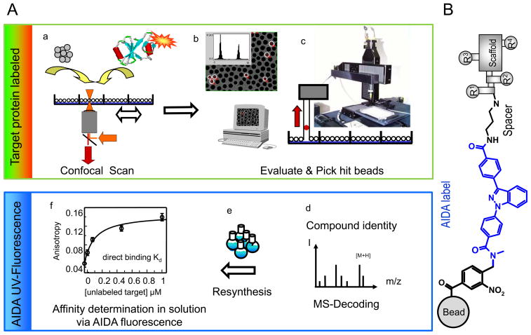
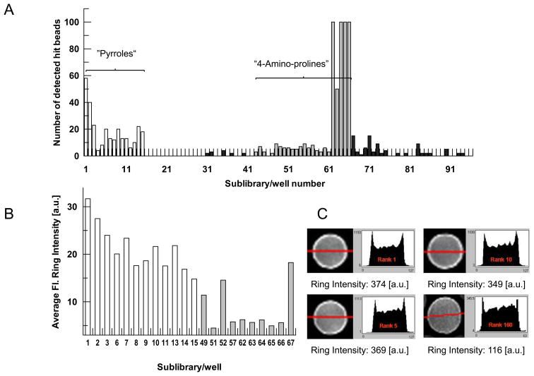
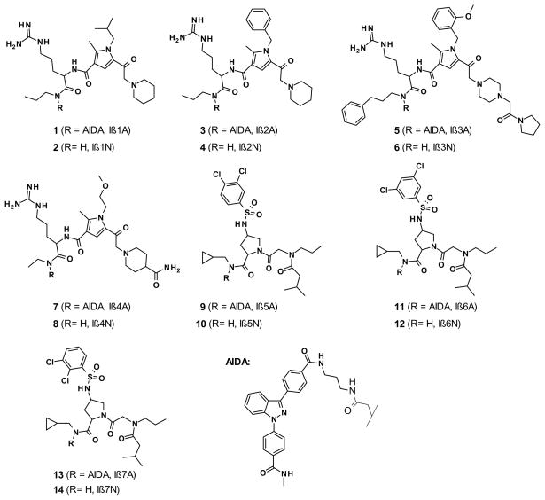
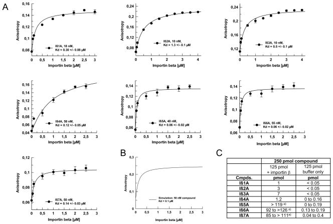
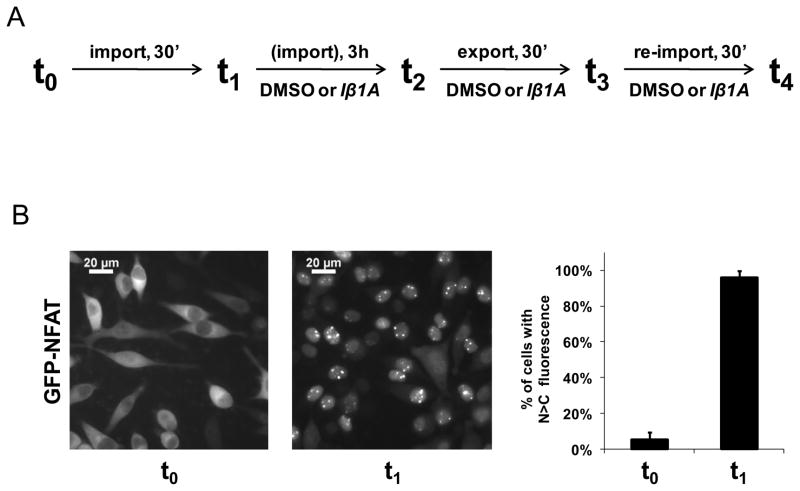
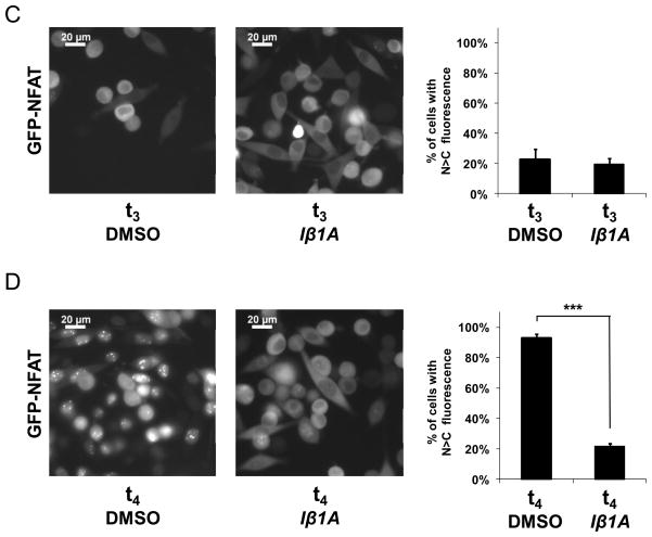
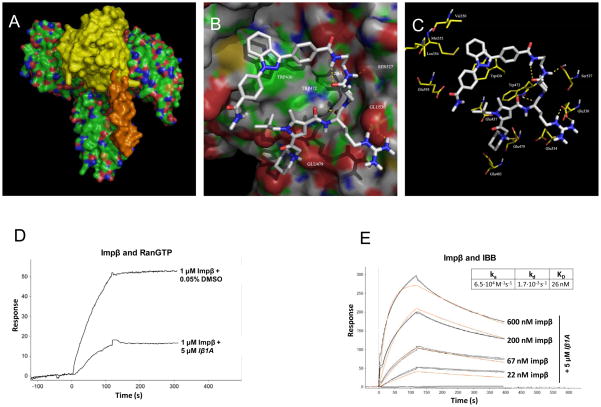
In eukaryotic cells, proteins and RNAs are transported between the nucleus and the cytoplasm by nuclear import and export receptors. Over the past decade, small molecules that inhibit the nuclear export receptor CRM1 have been identified, most notably leptomycin B. However, up to now no small molecule inhibitors of nuclear import have been described. Here we have used our automated confocal nanoscanning and bead picking method (CONA) for on-bead screening of a one-bead one-compound library to identify the first such import inhibitor, karyostatin 1A. Karyostatin 1A binds importin β with high nanomolar affinity and specifically inhibits importin α/β mediated nuclear import at low micromolar concentrations in vitro and in living cells, without perturbing transportin mediated nuclear import or CRM1 mediated nuclear export. Surface plasmon resonance binding experiments suggest that karyostatin 1A acts by disrupting the interaction between importin β and the GTPase Ran. As a selective inhibitor of the importin α/β import pathway, karyostatin 1A will provide a valuable tool for future studies of nucleocytoplasmic trafficking.


Figures

Figure 1
Figure 1. Affinity based screening of AIDA-tagged one-bead one-compound libraries by Confocal Nanoscanning (CONA) and fluorescence based secondary assays
A) The screening process is performed in six steps (a–f): a) starting with distribution of 1 mg of resin from each AIDA-tagged sublibrary into the wells of a 96-well microtiter plate, followed by incubation with fluorescently tagged target protein. b) Automated confocal nanoscanning (CONA) identifies relevant hit-beads where the target protein has bound to compounds on the bead surface. c) Hit beads are isolated by the bead-picking device of the CONA screening instruments and the compounds are cleaved from the resin. d) MS-analysis of the hit-compounds allows structure assignment for each hit bead. e) The identified hits are re-synthesized in mg quantities with and without the AIDA tracer. f) The fluorescence from the UV-dye AIDA is used in a generic secondary assay to quantify the affinity of the hit-compounds for the target protein and for compound ranking. Note: This process falls into two phases: a phase where the green-red fluorescence on the target protein is first used to identify hits by on-bead screening (green box) and a second phase where the UV-signal from the tracer molecule is used quantify the obtained primary hits in a generic secondary assay (blue box). B) Library set-up: one-bead one-compound libraries were synthesized on 90 μm TentaGel beads, using a photo-cleavable linker as attachment site (black), followed by a chemically robust UV-tracer “AIDA” (blue), a 3-carbon atom spacer (black) and the actual screening compound. The screening compounds are built around a central scaffold, decorated with four combinatorial sites. According to the split-mix-and-divide synthesis protocol used, the last two combinatorial sites are identical for each compound in any one sublibrary.
Figure 2
Figure 2. Primary on-bead screening analysis
A) Distribution of the number of hit-beads over one 96-well screening plate containing a diversity optimized subset of AIDA-tagged one-bead one-compound libraries. Each well number represents one sublibrary. B) Quantitative analysis of relative fluorescence ring intensities of hit-beads from pyrrole and amino-proline containing wells. Fluorescence ring intensity, is the quantitative parameter, indicating the amount of fluorescently tagged target protein which has bound to the bead-immobilized compounds. C) Four exemplary bead images with their corresponding fluorescence ring intensities and ranking.
Figure 3
Figure 3. Re-synthesized CONA derived hit compounds for importin β
Based on the MS-analysis of the individual hit-compounds and the building block-frequency analysis seven hit-compounds were selected for re-synthesis with (Iβ1A to Iβ7A) and without the AIDA tag (Iβ1N to Iβ7N) for further investigations in follow-up assays.
Figure 4
Figure 4. Determination of binding affinities (Kds) of hit compounds Iβ1A to Iβ7A for importin β
A) Anisotropy measurements using the AIDA-derived fluorescence signal were carried out with increasing concentrations of importin β. The resulting titration data was fitted to a 1:1 interaction model. B) Simulation of expected start- and end-anisotropy values for a ligand with a molecular weight of 1,000 Da and a globular shaped protein of 100 kDa, using the Perrin equation. C) HPLC-quantified recoveries of AIDA-tagged hit compounds after size-exclusion chromatography experiments in presence and absence of a saturating amount of importin β.
Figure 5
Figure 5. Effect of compounds Iβ1A to Iβ4A on in vitro nuclear import using permeabilized cells
Importin α/β mediated in vitro nuclear import using recombinant transport factors. Transport factors were added to permeabilized HeLa suspension cells together with cargo, energy and compounds. DMSO concentration was kept at 1% across the samples. Following a 30-minute reaction, nuclear fluorescence was analyzed by flow cytometry. The data points represent the average of 3 to 7 independent experiments. A. Compounds Iβ1A to Iβ7A and Iβ1N to Iβ7N were added at 10 μM final concentration together with FITC-BSA-NLS cargo and recombinantly expressed transport factors. B. Compounds Iβ1A to Iβ3A were added at 10 μM, 3.3 μM and 1 μM concentrations with FITC-BSA-NLS cargo and recombinantly expressed transport factors. C. Compounds Iβ1A to Iβ4A were added at 10 μM final concentration together with FITC-BSA-NLS cargo and cytosol as a source of nuclear transport factors. WGA: wheat germ agglutinin. D. Nuclear import assay with FITC-BSA-NLS cargo and recombinantly expressed transport factors. Compounds Iβ1A to Iβ4A were added at 10 μM final concentration only after the nuclear import reactions were terminated by hexokinase/glucose, followed by 30 minutes of incubation at 30°C to test for the loss of intranuclear FITC-BSA-NLS. E. Transportin mediated nuclear import using recombinant transport factors. Compounds Iβ1A to Iβ4A were added at 10 μM final concentration to permeabilized HeLa suspension cells together with FITC-M9-nucleoplasmin cargo.
Figure 6
Figure 6. Effect of Iβ1A on nucleocytoplasmic export and import of GFP-NFAT in living cells
A. Schematic representation of the experimental timeline to test the in vivo effect of Iβ1A on nuclear export and import of GFP-NFAT in HeLa cells. “Import” or “re-import” denotes conditions where nuclear import of GFP-NFAT is triggered with the addition of 1 μM ionomycin to the cells. “Export” denotes conditions where the ionomycin is washed out with cell culture medium. B. Cytoplasmic localization of GFP-NFAT from untreated, stably transfected HeLa cells and its nuclear translocation 30 minutes after inducing import. C. Localization of GFP-NFAT in the presence of either 0.25% DMSO or 25 μM Iβ1A 30 minutes after nuclear export was induced. A three-hour incubation period with DMSO or Iβ1A in the presence of ionomycin preceded the initiation of export. D. Localization of GFP-NFAT in the presence of either 0.25% DMSO or 25 μM Iβ1A 30 minutes after nuclear re-import of GFP-NFAT was induced with ionomycin on the same cells that underwent export in C. Images in B., C. and D. were collected of HeLa cells expressing GFP-NFAT by fluorescence microscopy. The graphs depict the percentage of cells with predominantly nuclear GFP-NFAT, as determined by visual inspection of at least 200 cells for each condition.
Figure 7
Figure 7. Molecular modeling and surface plasmon resonance measurements of the Iβ1A-importin beta interaction
A. Interaction between importin β (green, coloured by atom type), importin α (orange) and Ran-GTP (yellow). B. Docked structure of Iβ1A within its identified binding site Importin β as obtained by fully flexible protein-ligand docking using the program RosettaLigand (http://www.rosettacommons.org/). C. Pharmacophore model derived from fully flexible molecular docking studies. D. Binding of importin β at 1 μM concentration to GST-RanQ69LGTP non-covalently immobilized to a GST antibody chip in the presence of 5 μM Iβ1A or 0.05% DMSO. Binding of importin β at various concentrations in the presence of 5 μM Iβ1A to GST-IBB (E.) non-covalently immobilized to a GST antibody chip. Black lines represent actual data collected in duplicates and red lines are theoretical simulations derived from global fit on the dataset.

References

    1. Terry LJ, Shows EB, Wente SR. Crossing the nuclear envelope: hierarchical regulation of nucleocytoplasmic transport. Science. 2007;318:1412–1416. - PubMed
    1. Stewart M. Molecular mechanism of the nuclear protein import cycle. Nat Rev Mol Cell Biol. 2007;8:195–208. - PubMed
    1. Weis K. Importins and exportins: how to get in and out of the nucleus. Trends Biochem Sci. 1998;23:235. - PubMed
    1. Cook A, Bono F, Jinek M, Conti E. Structural biology of nucleocytoplasmic transport. Annu Rev Biochem. 2007;76:647–671. - PubMed
    1. Bednenko J, Cingolani G, Gerace L. Nucleocytoplasmic transport: navigating the channel. Traffic. 2003;4:127–135. - PubMed

Publication types

MeSH terms