Case Reports
. 2011 Jan;20(1):11-7.
doi: 10.1089/scd.2010.0139. Epub 2010 Oct 12.

Human Barrett's adenocarcinoma of the esophagus, associated myofibroblasts, and endothelium can arise from bone marrow-derived cells after allogeneic stem cell transplant

Affiliations
Case Reports

Human Barrett's adenocarcinoma of the esophagus, associated myofibroblasts, and endothelium can arise from bone marrow-derived cells after allogeneic stem cell transplant

Lloyd Hutchinson et al. Stem Cells Dev. 2011 Jan.

Abstract

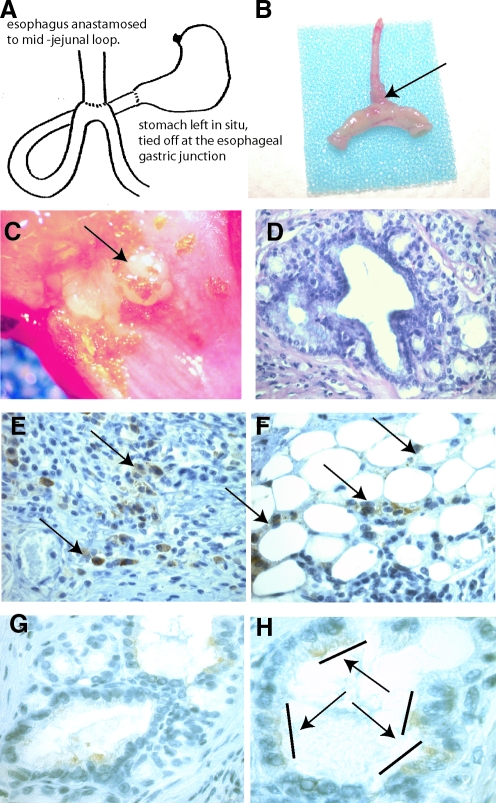
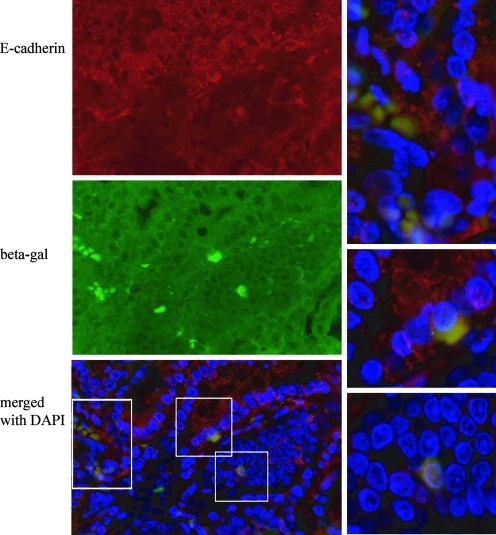
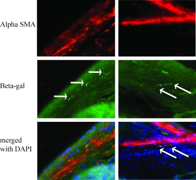
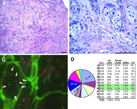
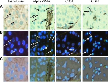
This study characterizes the contribution of bone marrow-derived cells (BMDCs) to Barrett's adenocarcinoma of the esophagus using a mouse surgical model of disease and human specimens. Transplantation of bone marrow expressing beta galactosidase into a wild-type mouse, followed by surgical esophagojejunostomy, allowed tracking of BMDCs into the surgical anastomosis and resulting Barrett's metaplasia. Human tissue from a male patient who had been transplanted with female bone marrow and later developed esophageal adenocarcinoma allowed us to tract donor-derived cells into the tumor. Using a combination of antibodies directed against beta-galactosidase (animal studies) and X/Y fluorescent in situ hybridization (FISH) (human studies), combined with specific lineage staining directed against epithelial, fibroblast, endothelial, and leukocyte markers, we show that bone marrow cells contribute to both the epithelial and stromal component of esophageal adenocarcinoma. These findings demonstrate that BMDCs can generate cancer-associated fibroblasts as well as contribute directly to epithelial cells in cancer of the esophagus.


Figures

FIG. 1.
FIG. 1.
Surgical mouse model of Barrett's metaplasia. (A) Cartoon depiction of the surgical esophagojejunostomy. (B) Actual excised esophagus and anastomosed jejunum from a mouse at 20 weeks. Arrow points to the thickened esophagus proximal to the anastomosis. (C) Opening of the surgical specimen reveals nodular thickened area proximal to the anastomosis, not involving the suture line. (D) H&E staining of a histological section through the anastomosis. Poorly organized glands lined by columnar epithelium with interspersed stromal tissue surrounded by squamous epithelial cells. Immunohistochemical staining directed against beta-galactosidase (brown cytoplasmic staining) reveals bone marrow cells within (E) inflammatory infiltrate (arrows). (F) Adipose tissue surrounding tumor (arrows) and (G, H) within gland structures. Bars and arrows show 2 and 3 contiguous cells that are marrow derived.
FIG. 2.
FIG. 2.
Epithelial cells of Barrett's metaplasia in the mouse are marrow derived. Immunofluorescence staining of tissue sections through the lower esophagus. E-cadherin (red), beta-galactosidase (green), and nuclei (blue) as labeled (40 ×). Merged images of bone marrow-derived cells expressing E-cadherin that are boxed are shown at higher magnification (60 ×).
FIG. 3.
FIG. 3.
Stromal cells within Barrett's metaplasia in the mouse are marrow derived. Immunofluorescence staining of tissue sections through the lower esophagus. Alpha smooth muscle actin (alpha SMA) (red), beta-galactosidase (green), and nuclei (blue) as labeled (40×). Arrows highlight bone marrow-derived cells expressing alpha SMA and beta galactosidase.
FIG. 4.
FIG. 4.
Human esophageal tumor arising in a transplant patient. (A) H&E-stained sections show a poorly differentiated adenosquamous carcinoma composed of islands and small clusters of tumor cells within a dense desmoplastic stroma; 10× magnification, scale bar = 100 μm. (B) On higher power the tumor shows mixed features of adenocarcinoma and squamous cell carcinoma with high-grade pleomorphic nuclei and mitotic activity. A reactive myofibroblastic stromal component and vessels are evident within the tumor; 40× magnification, scale bar = 100 μm. (C) Fluor-escent in situ hybridization (FISH) for X (green) and Y (red) chromosomes. A cluster of XX female tumor cells can be seen surrounded by stroma (arrows). (D) Enumeration of XY chromosome counts are shown in pie chart form and table.
FIG. 5.
FIG. 5.
Specific immunohistochemistry and X/Y FISH analysis performed in tandem on the same slide. (A) Immunohistochemistry as indicated (B) X (green) and Y (red) FISH followed by (C) an overlay for each grouping. Epithelial cells stained with anti-E-cadherin antibody and fibroblast cells within the stroma are stained with antibody against alpha smooth muscle actin; endothelial cells are stained with antibody directed against CD31 and leukocytes are stained with anti-CD45. Arrows point out 1X0Y and 2X0Y cells.

References

    1. Jiang Y. Jahagirdar BN. Reinhardt RL. Schwartz RE. Keene CD. Gonzalez Ortiz-XR. Reyes M. Lenvik T. Lund T. Blackstad M. Du J. Aldrich S. Lisberg A. Low WC. Largaespada DA. Verfaillie CM. Pluripotency of mesenchymal stem cells derived from adult marrow. Nature. 2002;418:41–46. - PubMed
    1. Krause DS. Theise ND. Collector MI. Henegariu O. Hwang S. Gardner R. Neutzel S. Sharkis SJ. Multi-organ, multi-lineage engraftment by a single bone marrow-derived stem cell. Cell. 2002;105:369–377. - PubMed
    1. Wagers AJ. Sherwood RI. Christensen JL. Weissman IL. Little evidence for developmental plasticity of adult hematopoietic stem cells. Science. 2002;297:2256–2259. - PubMed
    1. Brittan M. Chance V. Elia G. Poulsom R. Alison MR. MacDonald TT. Wright NA. A regenerative role for bone marrow following experimental colitis: contribution to neovasculogenesis and myofibroblasts. Gastroenterol. 2005;128:1984–1995. - PubMed
    1. Sasaki M. Abe R. Fujita Y. Ando S. Inokuma D. Shimizu H. Mesenchymal stem cells are recruited into wounded skin and contribute to wound repair by transdifferentiation into multiple skin cell type. J Immunol. 2008;180:2581–2587. - PubMed

Publication types

MeSH terms