. 2010 Dec;12(12):1792-808.
doi: 10.1111/j.1462-5822.2010.01509.x.

Toxoplasma gondii protease TgSUB1 is required for cell surface processing of micronemal adhesive complexes and efficient adhesion of tachyzoites

Affiliations

Toxoplasma gondii protease TgSUB1 is required for cell surface processing of micronemal adhesive complexes and efficient adhesion of tachyzoites

Vanessa Lagal et al. Cell Microbiol. 2010 Dec.

Abstract

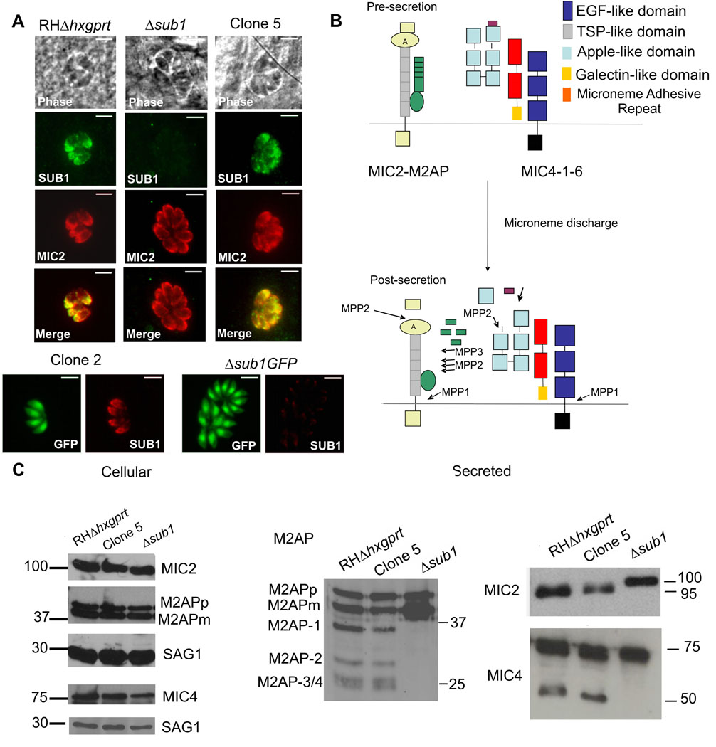
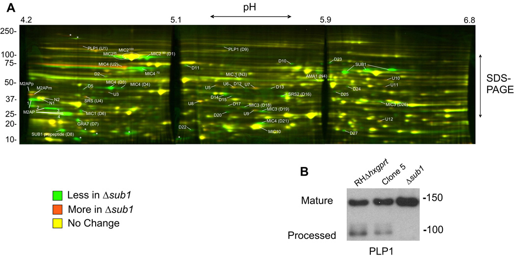
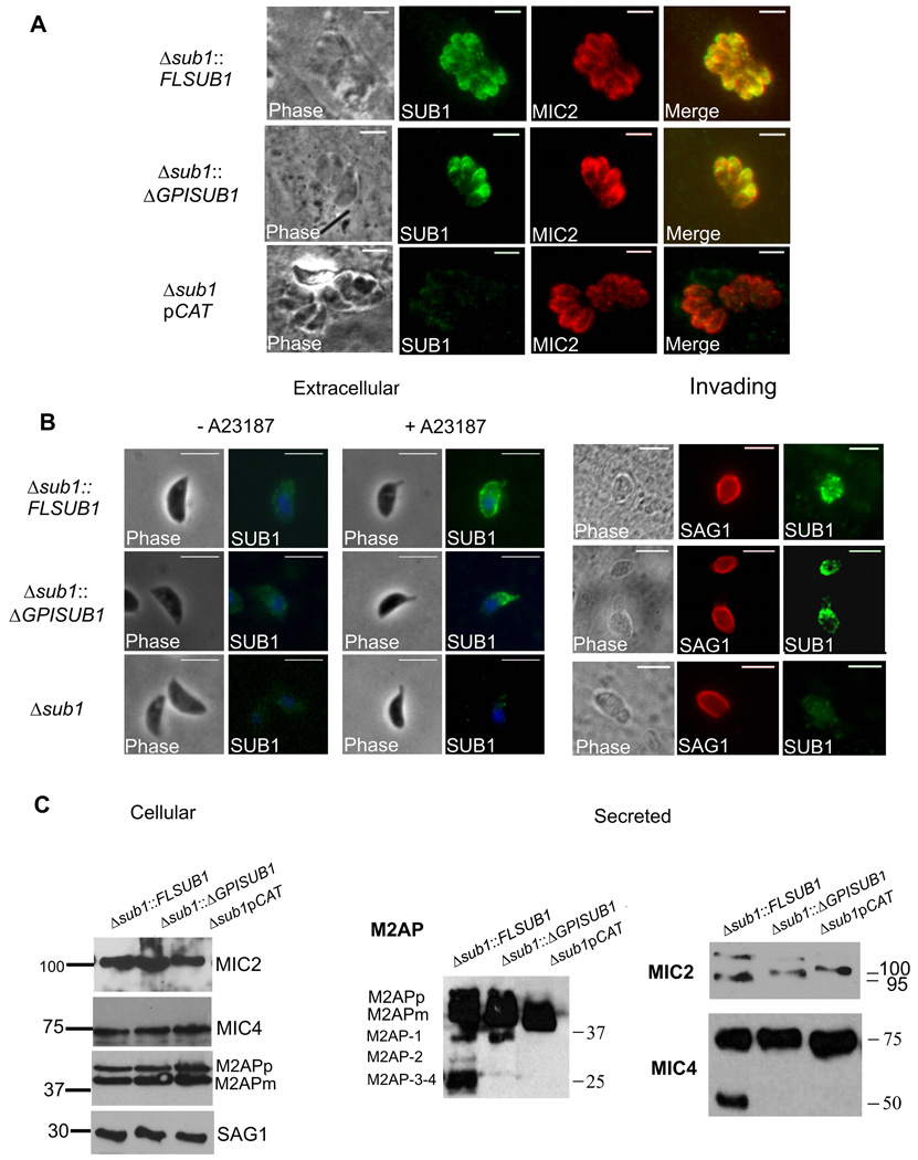
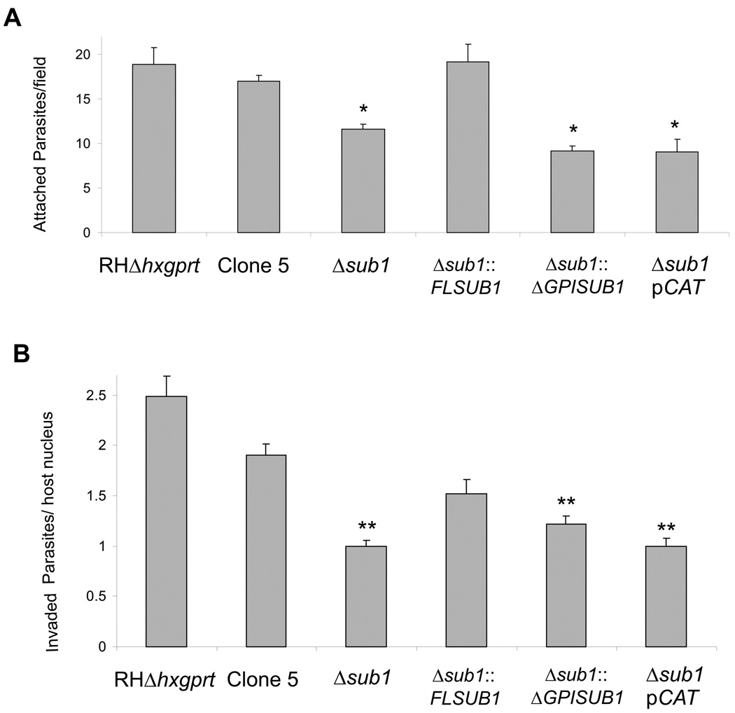
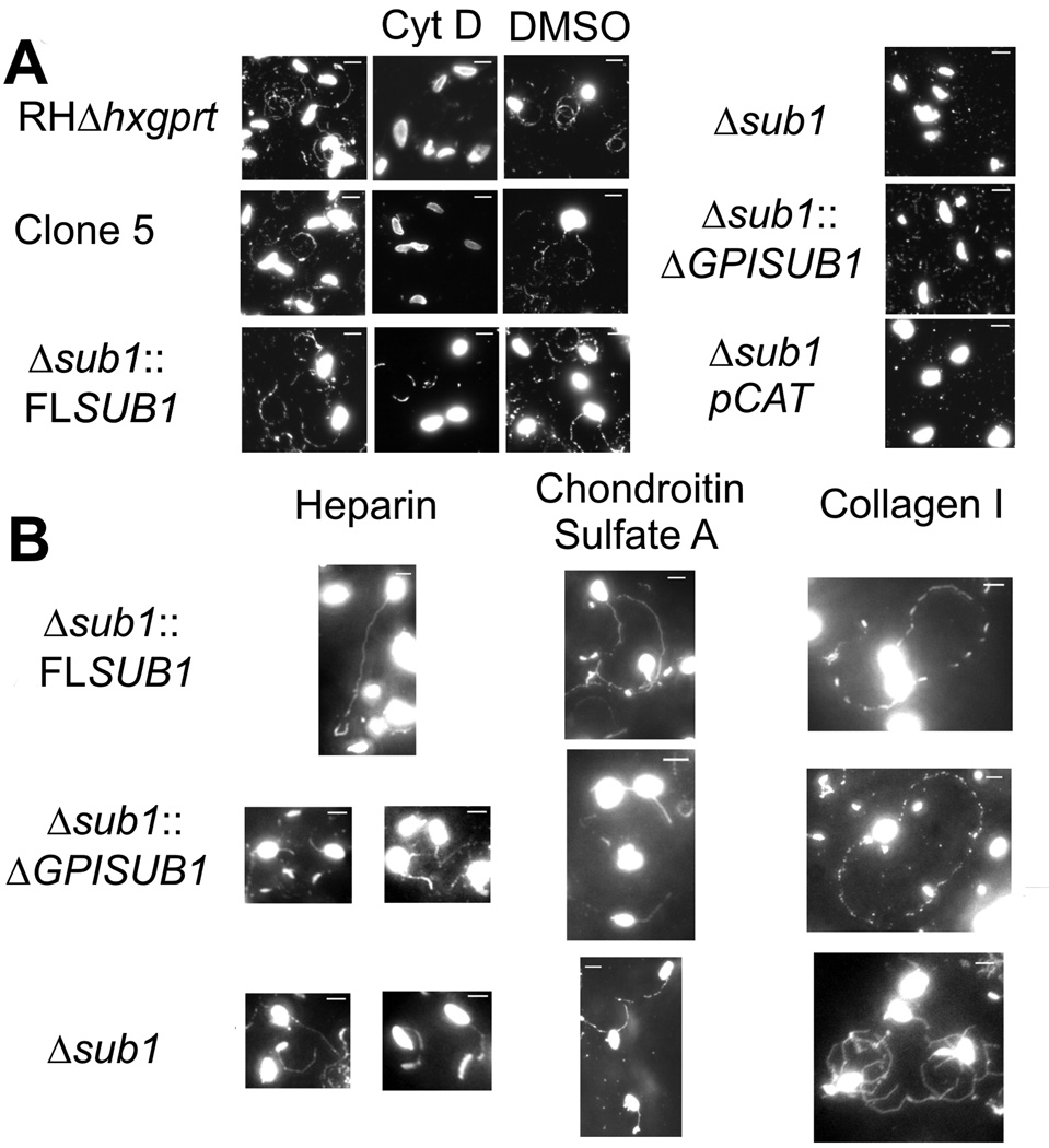
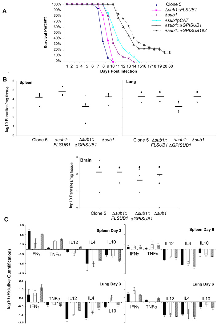
Host cell invasion by Toxoplasma gondii is critically dependent upon adhesive proteins secreted from the micronemes. Proteolytic trimming of microneme contents occurs rapidly after their secretion onto the parasite surface and is proposed to regulate adhesive complex activation to enhance binding to host cell receptors. However, the proteases responsible and their exact function are still unknown. In this report, we show that T. gondii tachyzoites lacking the microneme subtilisin protease TgSUB1 have a profound defect in surface processing of secreted microneme proteins. Notably parasites lack protease activity responsible for proteolytic trimming of MIC2, MIC4 and M2AP after release onto the parasite surface. Although complementation with full-length TgSUB1 restores processing, complementation of Δsub1 parasites with TgSUB1 lacking the GPI anchor (Δsub1::ΔGPISUB1) only partially restores microneme protein processing. Loss of TgSUB1 decreases cell attachment and in vitro gliding efficiency leading to lower initial rates of invasion. Δsub1 and Δsub1::ΔGPISUB1 parasites are also less virulent in mice. Thus TgSUB1 is involved in micronemal protein processing and regulation of adhesive properties of macromolecular adhesive complexes involved in host cell invasion.


Figures

Figure 1
Figure 1. TgSUB1 disruption alters MIC proteolytic processing
(A) Immunofluorescence of Δsub1 strains and sibling clones (Clone 2 for Δsub1 GFP; Clone 5 for Δsub1). Controls include the wild-type RH GFP-Δhxgprt and RHΔhxgprt strains. Siblings correspond to wild-type parasites stably transfected with the selectable marker but without TgSUB1 disruption. TgSUB1 is labeled with the TgSUB1 AE653 rabbit antiserum. MIC2 is labeled with the mouse mAb 6D10 and was used as a marker for microneme localization. Scale bar is 5 µm (B) Schematic representation of the cell surface proteolytic trimming of the micronemal adhesive complexes MIC2-M2AP and MIC1-4–6. “A” denotes the A-domain of MIC2 protein. Microneme Adhesive Repeat refers to the specialized MIC1 domain which discriminates between glycan residues (Blumenschein et al., 2007). Activity of the proteases MPP1-2–3 is represented. Because the first step in M2AP processing is not inhibited by protease inhibitors that inhibit MPP2 (ALLM, ALLN), a second protease activity termed MPP3 was proposed (Zhou et al., 2004). The MPP1 MIC cleavage sites are consistent with MPP1 being a rhomboid serine protease such as the parasite surface rhomboids TgROM 4 or TgROM5 (Brossier et al., 2005, Dowse et al., 2005, Buguliskis et al., 2010). (C) Western Blot analysis of the MIC2-4 and M2AP cellular and secreted products. Blots were probed with appropriate antibodies as described in experimental procedures. Molecular standards are in kDa. The rabbit anti-MIC4 antibody used was raised against the A1 and A2 apple domains, and recognizes the 72 and 70 kDa precursor forms and the 50 kDa secreted product but not the 15 kDa secreted product. M2APp: precursor form; M2APm: mature form.
Figure 2
Figure 2. 2D-DIGE analysis of microneme secreted products
(A) Secreted products of the Δsub1GFP and Clone 2 strains were covalently labeled with Cy3 (green) or Cy5 (red) respectively, and separated by a two-dimension gel electrophoresis (pH 4.2–6.8 first dimension and 12.5% SDS-PAGE second dimension). Protein spots that are diminished or absent in the Δsub1 secreted sample compared to the Clone 2 (a sibling transfectant that still expresses TgSUB1), are depicted as green spots while proteins that are increased are depicted as orange spots. Yellow spots depict proteins that do not change significantly in abundance between the two strains. Marked proteins were excised and identified by mass spectrometry analysis. The asterisks indicate spots not sufficiently visible on the Coomassie gel to excise. (B) Western Blot analysis of the PLP1 secreted products. PLP1 was detected with appropriate antibody. Molecular weights are in kDa.
Figure 3
Figure 3. Complementation of the MIC surface processing by TgSUB1 complemented Δsub1 strains
(A) Immunofluorescence of TgSUB1 complemented Δsub1 strains. All strains were simultaneously labeled with TgSUB1 antiserum AE653 and MIC2 monoclonal antibody 6D10, a marker for micronemes. Scale bar is 5 µm (B) Surface detection of TgSUB1 in the 3 representative strains: Δsub1::FLSUB1, Δsub1::ΔGPISUB1 and Δsub1. Extracellular parasites were treated with the ionophore A23187 to induce microneme discharge. Tachyzoite nuclei were stained with DAPI and TgSUB1 was immunolabeled with the TgSUB1-specific antibody AE653 without cellular permeabilization. Freshly egressed parasites were inoculated onto HFF monolayers and invading tachyzoites were immunolabeled with TgSUB1 AE653 antiserum and the mouse anti-SAG1 DG52 monoclonal antibody without permeabilization. Scale bar is 5 µm (C) Western Blot analysis of the MIC2, MIC4 and M2AP cellular and secreted products collected from the Δsub1::FLSUB1, Δsub1::ΔGPISUB1 and Δsub1pCAT strains. For MIC2 some cellular MIC2 is evident in addition to secreted MIC2 in the ESA from the 2 complemented lines (migrating just above 100 kDa). Blots were probed with appropriate antibodies as described in Experimental Procedures. Molecular weights are in kDa. M2APp: precursor form; M2APm: mature form.
Figure 4
Figure 4. Attachment and invasion phenotypes of strains lacking TgSUB1-dependent MIC cell surface processing
(A) Quantification of attachment to glutaraldehyde-fixed HFF cells expressed as the number of attached parasites counted per field. An asterisk indicates a statistically significant difference compared with RHΔhxgprt, Clone 5 and Δsub1::FLSUB1 (P<0.05, two tailed Student’s t test) (B) Quantification of 15 minute invasion assays expressed in number of invaded parasites per host cell counted. A double asterisk indicates a statistically significant difference compared with RHΔhxgprt and Clone 5 (P<0.05, two tailed Student’s t test). Data are mean values ± SEM.
Figure 5
Figure 5. Motility of strains lacking TgSUB1 assessed by trail deposition
All strains were allowed to glide on glass coverslips coated with FBS (A) or Heparin, Chondroitin sulfate A and Collagen I (B). Trails deposited by gliding parasites were revealed with the mouse anti-SAG1 monoclonal antibody DG52 and visualized by immunofluorescence microscopy. Parasites were also pretreated with the gliding inhibitor cytochalasin D (cytD) as negative control or with DMSO (solvent control). Scale bar is 5 µm.
Figure 6
Figure 6. Strains lacking TgSUB1 have decreased virulence in the mouse model
250 tachyzoites were injected intravenously into groups of 5 female CD-1 mice. (A) Mouse survival was monitored daily over a period of 8 weeks. The Δsub1 and the Δsub1pCAT strains are less virulent than the Clone 5 and the Δsub1::FLSUB1 strains (P <0.05). The Δsub1::ΔGPISUB1 was significantly less virulent than the Δsub1::FLSUB1 and Δsub1 strains (P<0.001). Similar results were obtained with a second independent Δsub1::ΔGPISUB1 clone (Δsub1:: ΔGPISUB #2). Data are pooled from 3 independent experiments (at least 15 mice total). (B) Parasite tissue burden after 6 days of infection with the Clone 5, Δsub1, Δsub1::FLSUB1 and Δsub1::ΔGPISUB1 strains. Spleen and lung parasite burdens were lower for Δsub1::ΔGPISUB1 compared to Clone 5, Δsub1, and Δsub1::FLSUB1 strains, but data were statistically significant for the lung only (two-tailed Student’s t-test, P< 0.05). No significant differences were observed in the brain where fewer parasites were detected for all strains. Data are pooled from 2 independent experiments and show burdens for each mouse tested with average value represented by the dark line (C) Mice cytokine tissue levels after 3 and 6 days of infection with Clone 5 (black bars), Δsub1::ΔGPISUB1 (white bars) and Δsub1 (gray bars) strains. Significant differences in the cytokine levels were observed at day 3: Spleen: for TNFα between the clone 5 strain compared to the Δsub1 and Δsub1::ΔGPISUB1 strains. Lung: for IL12 and IL4 cytokines between the three strains and for IL10 between the Δsub1::ΔGPISUB1 strain compared to the clone 5 and Δsub1 strains. At day 6, no significant differences in the cytokine levels were observed except for TNFα between the Δsub1::ΔGPISUB1 strain compared to the Δsub1 and clone 5 strains (two-tailed Student’s t-test, P< 0.05).

Similar articles

Cited by

References

    1. Barragan A, Brossier F, Sibley DL. Transepithelial migration of toxoplasma gondii involves an interaction of intercellular adhesion molecule 1 (ICAM-1) with the parasite adhesin MIC2. Cell. Microbiol. 2005;7:561–568. - PubMed
    1. Barragan A, Sibley DL. Transepithelial migration of toxoplasma gondii is linked to parasite motility and virulence. J. Exp. Med. 2002;195:1625–1633. - PMC - PubMed
    1. Baum J, Papenfuss A, Baum B, Speed T, Cowman A. Regulation of apicomplexan actin-based motility. Nat. Rev. Microbiol. 2006;4:621–628. - PubMed
    1. Binder EM, Lagal V, Kim K. The prodomain of Toxoplasma gondii GPI-anchored subtilase TgSUB1 mediates its targeting to micronemes. Traffic. 2008;9:1485–1496. - PMC - PubMed
    1. Black M, Arrizabalaga G, Boothroyd J. Ionophore-resistant mutants of Toxoplasma gondii reveal host cell permeabilization as an early event in egress. Mol. Cell. Biol. 2000;20:9399–9408. - PMC - PubMed

Publication types

MeSH terms