. 2010 Oct 15;346(2):237-46.
doi: 10.1016/j.ydbio.2010.07.029. Epub 2010 Aug 1.

Left-right patterning in the mouse requires Epb4.1l5-dependent morphogenesis of the node and midline

Affiliations

Left-right patterning in the mouse requires Epb4.1l5-dependent morphogenesis of the node and midline

Jeffrey D Lee et al. Dev Biol. .

Abstract

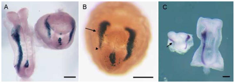
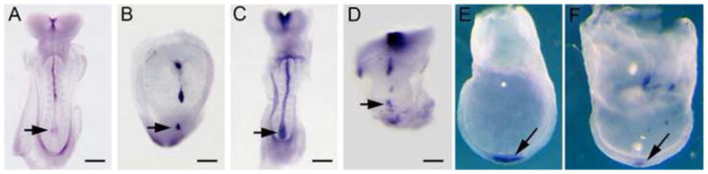
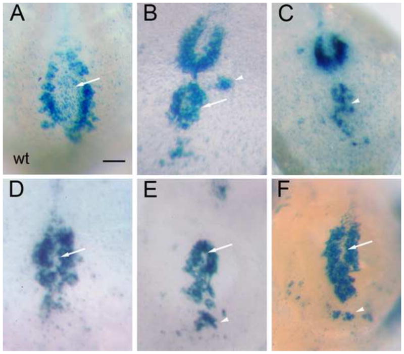
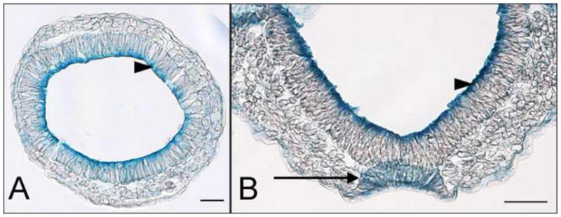
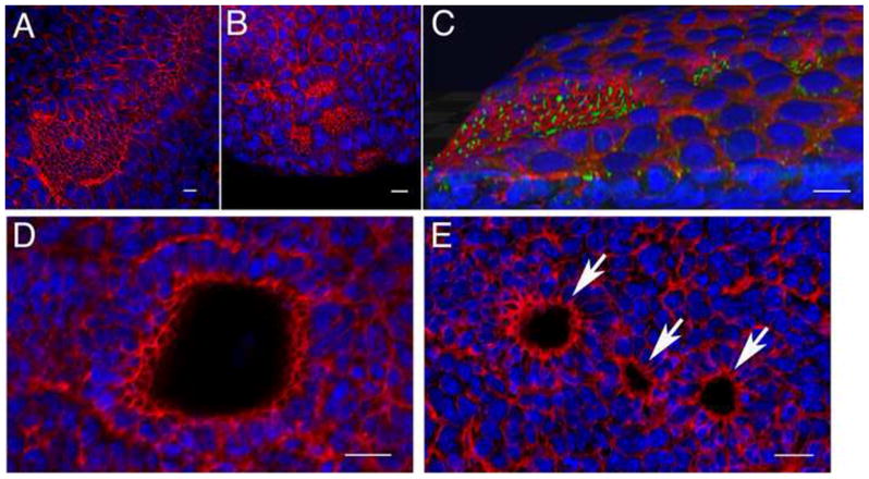
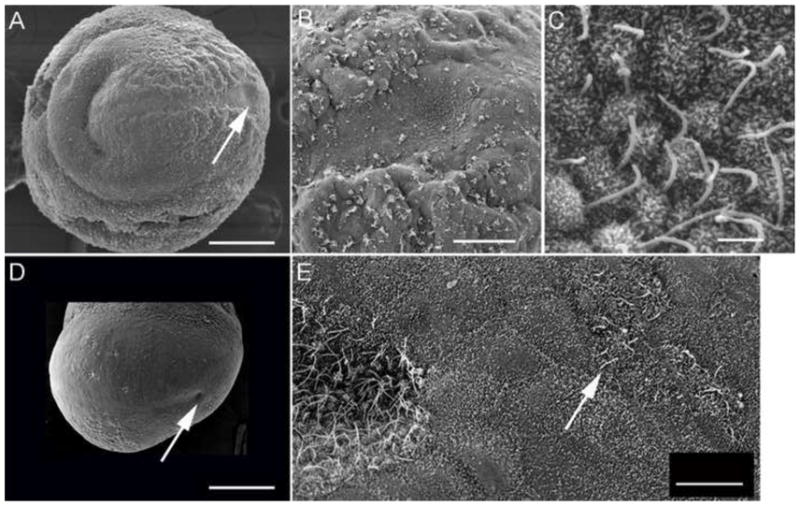
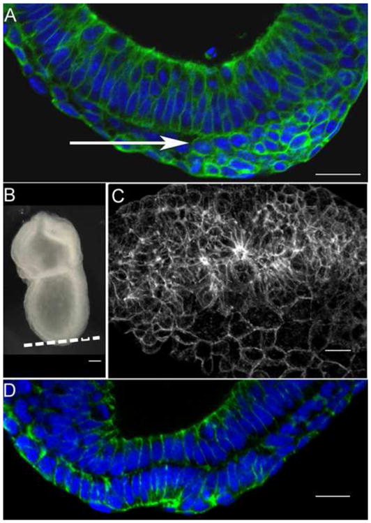
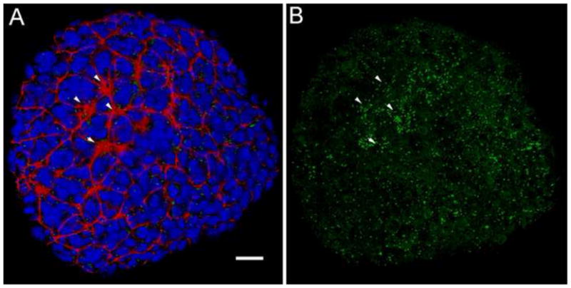
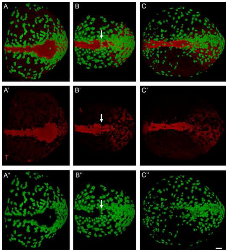
The mouse node is a transient early embryonic structure that is required for left-right asymmetry and for generation of the axial midline, which patterns neural and mesodermal tissues. The node is a shallow teardrop-shaped pit that sits at the distal tip of the early headfold (e7.75) embryo. The shape of the node is believed to be important for generation of the coherent leftward fluid flow required for initiation of left-right asymmetry, but little is known about the morphogenesis of the node. Here we show that the FERM domain protein Lulu/Epb4.1l5 is required for left-right asymmetry in the early mouse embryo. Unlike other genes previously shown to be required for left-right asymmetry in the mouse, lulu is not required for specification of node cell identity, for Nodal signaling in the node or for ciliogenesis. Instead, lulu is required for proper morphogenesis of the node and midline. The precursors of the wild-type node undergo a series of rapid morphological transitions. First, node precursors arise from an epithelial-to-mesenchymal transition at the anterior primitive streak. While in the mesenchymal layer, the node precursors form several ciliated rosette-like clusters; they then rapidly undergo a mesenchymal-to-epithelial transition to insert into the outer, endodermal layer of the embryo. In lulu mutants, node precursor cells are specified and form clusters, but those clusters fail to coalesce to make a single continuous node epithelium. The data suggest that the assembly of the contiguous node epithelium from mesenchymal clusters requires a rapid reorganization of apical-basal polarity that depends on Lulu/Epb4.1l5.


Figures

Fig. 1
Fig. 1
Left-right asymmetry is disrupted in lulu mutant embryos. (A) Dorsal views of nodal-lacZ expression in e8.5 wild-type (left) and lulu mutant embryos (right). Nodal-lacZ is expressed in the left lateral plate mesoderm (LPM) in wild type, but is expressed bilaterally in the mutant. (B) Ventral view of another lulu mutant embryo, showing Nodal-lacZ expression in both the left and right (arrow) LPM. The expression in the right LPM does not extend as far posteriorly (arrowhead) as in the left LPM. Note the abnormal expression of nodal-lacZ in the region of the mutant node in (A) and (B). (C) lefty2 RNA expression is bilateral in lulu (left; ventral view) (seen in 3/3 mutant embryos examined), but is confined to the left LPM in wild type (right; dorsal view). As with Nodal-lacZ, lefty expression in the right LPM (arrow) does not extend as far posteriorly as in the left LPM. Scale bars = 150μm.
Fig. 2
Fig. 2
Abnormal expression of midline and node markers in lulu embryos, assayed by in situ hybridization. (A, B) Shh expression in e8.5 wild-type (A) and lulu (B) embryos. (C, D) FoxA2 expression in e8.5 wild-type (C) and lulu (D) embryos. Expression of both Shh and FoxA2 is discontinuous in the lulu midline. (E, F) FoxJ1 expression is expressed in a lateral view of an e7.5 wild-type (E) and a frontal view of a lulu (F) node arrows, but the expression level is lower in the mutant. Arrows point out the node. Scale bars = 150μm in A–D.
Fig. 3
Fig. 3
Variable disruption of node morphology in lulu mutant embryos, as revealed by the pattern of nodal-lacZ expression. Ventral views of wild type (A) and lulu nodes (B–F), in which node shape is outlined by the expression of nodal-lacZ in the crown cells. Anterior is to the top. Node shape is highly variable in the mutants; defects include loss or reduction of the node pit (arrows, D–F, compare to arrow in A), ectopic crown cells (arrowheads, B, C, E, F), and an apparently duplicated node (arrow, B). All images are at the same magnification; scale bar = 20 μm.
Fig. 4
Fig. 4
Expression of lulu in the node. (A, B) The LuluGT2-β-gal fusion protein (blue) is enriched apically in the epiblast epithelium (arrowheads) at both transverse sections of e7.5 embryos (A) and frontal sections of headfold stage embryos (B) that are heterozygous for the gene trap allele. Localization of the gene trap fusion protein parallels the localization of the endogenous Lulu/Epb4.1l5 protein (Lee et al., 2007). At the headfold stage (B), the epithelium of the node has assembled (arrow). The LuluGT2-β-gal fusion protein is present throughout these cells, rather than being enriched at the apical surface.
Fig. 5
Fig. 5
Abnormal cellular organization of the lulu node. Phalloidin (red) and DAPI (blue) staining revealed that multiple node pits form in lulu embryos. (A) Phalloidin staining of the distal surface of an e7.75 wild-type embryo shows the organization of the cells with small apical surfaces in the node and midline, compared to the large surfaces of the surrounding squamous cells of the endoderm. Anterior is to the upper right. (B) At the same stage in lulu embryos, cells with small apical surfaces are found in several separate clusters; this is an extreme example of the disruptions of the node in lulu embryos. Anterior is to the upper left. (C, D, E) Phalloidin: red. DAPI: blue. (C) 3D rendering of an e7.75 lulu embryo reveals that ectopic node cells form pits. Several clusters of cells with long cilia (marked by expression of Arl13b in green) are present outside the main node region; even the small clusters are depressed from the surface of the endoderm in small pits. Anterior to the left. See also Supplemental Movie 1. (D) A confocal section parallel to the surface of the wild-type node highlights the pit of the node, seen here as the empty circle surrounded by cells with small apical surfaces. (E) A similar confocal section through the node region of a lulu embryo shows three separate pits (arrows). Scale bars = 25μm.
Fig. 6
Fig. 6
Scanning electron micrographs of wild type (A, B, C) and lulu mutant (D, E) embryos, e7.75. Anterior is to the left in all panels. The node is located distally on the ventral surface of the embryo (arrow, A). A higher magnification view (B) shows the node is visible as a concave pit of cells with small apical surfaces, surrounded by squamous visceral endoderm cells; node cells have long cilia (C). (D) lulu mutant, lateral/ventral view. This node in this lulu embryo (arrow) is approximately half the diameter of the wild-type node, and is less deep. (D) A higher magnification view shows that cilia on the lulu node appear normal. Ectopic cells with long, node-like are found to the posterior of the node (arrow), surrounded by cells of the visceral endoderm. Scale bars in A, C = 200 μm; B = 50 μm; C = 2 μm; E = 10 μm.
Fig. 7
Fig. 7
Sequential epithelial-to-mesenchymal and mesenchymal-to-epithelial transitions produce the wild-type node. E-cadherin: green. DAPI: blue. (A) At the late streak stage, cells of the presumptive node accumulate at the anterior primitive streak between the epithelial layers of the epiblast and endoderm (arrow), after having delaminated from the epiblast layer (above the mesenchymal cells in this image). These cells express E-cadherin (green) around their circumferences. (B) A late streak embryo, indicating the plane of the optical section in (C). (C) E-cadherin expression (white) reveals that the presumptive node cells between the epiblast and endoderm layers form several large three-dimensional rosette-like structures in the presumptive node region, where E-cadherin is enriched at the center of each rosette. (D) Within a few hours, the cells of the presumptive node have completed the mesenchymal-to-epithelial transition. The node cells have incorporated into the outer layer of the embryo and express apical E-cadherin. The node has formed its characteristic pit at the distal tip of embryo. Scale bars in A, C, D = 25 μm; in D = 50 μm.
Fig. 8
Fig. 8
Groups of ciliated presumptive node cells are present below the endodermal layer in the wild-type embryo. (A, B) 3D rendering of a ventral view of an e7.25 wild-type embryo, early in the process of node morphogenesis. (A) Red = phalloidin; blue =DAPI; green = Arl13b. Several clusters of cells with small apical surfaces are visible on surface of the embryo (arrowheads). At this stage, the nuclei between the clusters belong to the visceral endoderm (Kwon et al., 2008). (B) Same embryo as in (A), Arl13b channel only, to visualize cilia. Clusters of long cilia are seen at the position of the clusters seen in phalloidin staining (arrowheads); additional cells with long cilia are found beneath the endoderm (compare to (A)). Note that short cilia are also broadly distributed on endoderm cells. Scale bar = 25 μm.
Fig. 9
Fig. 9
The organization of the node and midline is mirrored by the organization of cells of the visceral endoderm. Green = AFP-GFP, marks the visceral endoderm. Red = T; expressed in the node, axial midline and primitive streak. Anterior to the left; posterior (the primitive streak) to the right. (A) The axial midline of the wild-type headfold (e7.75) embryo. The formation of the node and midline is complete at this stage. Most visceral endoderm cells (green) have dispersed by this stage, except for those lying over the primitive streak (right). At this stage, a single row of visceral endoderm cells surrounds the node and midline. (A′) T channel only shows the shape of the node and midline. (A″) GFP channel shows the arrangement of visceral endoderm cells around the wild-type node and midline. (B and C) lulu mutant embryos; (B′ and C′) T channel only; (B″ and C″) GFP channel only. (B). In this lulu embryo the node appears to be pinched off into two adjacent clusters of node cells, and AFP+ cells align between the two node-like regions (arrow). (C) In this mutant, the node is reduced in size. A few AFP-GFP+ visceral endoderm cells are present over the axial midline of this mutant embryo and others lie over the posterior node. All images are at the same magnification; scale bar = 50 μm.

Similar articles

Cited by

References

    1. Amack JD, Wang X, Yost HJ. Two T-box genes play independent and cooperative roles to regulate morphogenesis of ciliated Kupffer’s vesicle in zebrafish. Dev Biol. 2007;310:196–210. - PubMed
    1. Ang SL, Rossant J. HNF–3β is essential for node and notochord formation in mouse development. Cell. 1994;78:561–74. - PubMed
    1. Basu B, Brueckner M. Cilia multifunctional organelles at the center of vertebrate left-right asymmetry. Curr Top Dev Biol. 2008;85:151–74. - PubMed
    1. Beckers A, Alten L, Viebahn C, Andre P, Gossler A. The mouse homeobox gene Noto regulates node morphogenesis, notochordal ciliogenesis, and left right patterning. Proc Natl Acad Sci USA. 2007;104:15765–70. - PMC - PubMed
    1. Beddington SP. An autoradiographic analysis of the potency of embryonic ectoderm in the 8th day postimplantation mouse embryo. J Embryol Exp Morphol. 1981;64:87–104. - PubMed

Publication types