HDAC inhibition attenuates inflammatory, hypertrophic, and hypertensive responses in spontaneously hypertensive rats
- PMID: 20679181
- PMCID: PMC2931819
- DOI: 10.1161/HYPERTENSIONAHA.110.154567
HDAC inhibition attenuates inflammatory, hypertrophic, and hypertensive responses in spontaneously hypertensive rats
Abstract
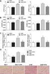
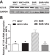
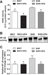
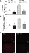
Reactive oxygen species and proinflammatory cytokines contribute to cardiovascular diseases. Inhibition of downstream transcription factors and gene modifiers of these components are key mediators of hypertensive response. Histone acetylases/deacetylases can modulate the gene expression of these hypertrophic and hypertensive components. Therefore, we hypothesized that long-term inhibition of histone deacetylase with valproic acid might attenuate hypertrophic and hypertensive responses by modulating reactive oxygen species and proinflammatory cytokines in SHR rats. Seven-week-old SHR and WKY rats were used in this study. Following baseline blood pressure measurement, rats were administered valproic acid in drinking water (0.71% wt/vol) or vehicle, with pressure measured weekly thereafter. Another set of rats were treated with hydralazine (25 mg/kg per day orally) to determine the pressure-independent effects of HDAC inhibition on hypertension. Following 20 weeks of treatment, heart function was measured using echocardiography, rats were euthanized, and heart tissue was collected for measurement of total reactive oxygen species, as well as proinflammatory cytokine, cardiac hypertrophic, and oxidative stress gene and protein expressions. Blood pressure, proinflammatory cytokines, hypertrophic markers, and reactive oxygen species were increased in SHR versus WKY rats. These changes were decreased in valproic acid-treated SHR rats, whereas hydralazine treatment only reduced blood pressure. These data indicate that long-term histone deacetylase inhibition, independent of the blood pressure response, reduces hypertrophic, proinflammatory, and hypertensive responses by decreasing reactive oxygen species and angiotensin II type1 receptor expression in the heart, demonstrating the importance of uncontrolled histone deacetylase activity in hypertension.
Figures


Similar articles
-
Lysine deacetylase inhibition attenuates hypertension and is accompanied by acetylation of mineralocorticoid receptor instead of histone acetylation in spontaneously hypertensive rats.Naunyn Schmiedebergs Arch Pharmacol. 2016 Jul;389(7):799-808. doi: 10.1007/s00210-016-1246-2. Epub 2016 Apr 22. Naunyn Schmiedebergs Arch Pharmacol. 2016. PMID: 27106211
-
Histone deacetylase inhibition attenuates cardiac hypertrophy and fibrosis through acetylation of mineralocorticoid receptor in spontaneously hypertensive rats.Mol Pharmacol. 2015 May;87(5):782-91. doi: 10.1124/mol.114.096974. Epub 2015 Feb 9. Mol Pharmacol. 2015. PMID: 25667225
-
Persistent cardiovascular effects of chronic renin-angiotensin system inhibition following withdrawal in adult spontaneously hypertensive rats.J Hypertens. 2001 Aug;19(8):1393-402. doi: 10.1097/00004872-200108000-00007. J Hypertens. 2001. PMID: 11518847
-
Therapeutic potential of the epidermal growth factor receptor transactivation in hypertension: a convergent signaling pathway of vascular tone, oxidative stress, and hypertrophic growth downstream of vasoactive G-protein-coupled receptors?Can J Physiol Pharmacol. 2007 Jan;85(1):97-104. doi: 10.1139/y06-097. Can J Physiol Pharmacol. 2007. PMID: 17487249 Review.
-
Three 4-letter words of hypertension-related cardiac hypertrophy: TRPC, mTOR, and HDAC.J Mol Cell Cardiol. 2011 Jun;50(6):964-71. doi: 10.1016/j.yjmcc.2011.02.004. Epub 2011 Feb 19. J Mol Cell Cardiol. 2011. PMID: 21320507 Free PMC article. Review.
Cited by
-
Mechanical regulation of epigenetics in vascular biology and pathobiology.J Cell Mol Med. 2013 Apr;17(4):437-48. doi: 10.1111/jcmm.12031. Epub 2013 Apr 3. J Cell Mol Med. 2013. PMID: 23551392 Free PMC article. Review.
-
Intestinal microbiota: A promising therapeutic target for hypertension.Front Cardiovasc Med. 2022 Nov 15;9:970036. doi: 10.3389/fcvm.2022.970036. eCollection 2022. Front Cardiovasc Med. 2022. PMID: 36457803 Free PMC article. Review.
-
Valproic acid: an anticonvulsant drug with potent antinociceptive and anti-inflammatory properties.Naunyn Schmiedebergs Arch Pharmacol. 2013 Jul;386(7):575-87. doi: 10.1007/s00210-013-0853-4. Epub 2013 Apr 14. Naunyn Schmiedebergs Arch Pharmacol. 2013. PMID: 23584602
-
Lysine deacetylase inhibition attenuates hypertension and is accompanied by acetylation of mineralocorticoid receptor instead of histone acetylation in spontaneously hypertensive rats.Naunyn Schmiedebergs Arch Pharmacol. 2016 Jul;389(7):799-808. doi: 10.1007/s00210-016-1246-2. Epub 2016 Apr 22. Naunyn Schmiedebergs Arch Pharmacol. 2016. PMID: 27106211
-
β-adrenergic Receptor Stimulation Revealed a Novel Regulatory Pathway via Suppressing Histone Deacetylase 3 to Induce Uncoupling Protein 1 Expression in Mice Beige Adipocyte.Int J Mol Sci. 2018 Aug 17;19(8):2436. doi: 10.3390/ijms19082436. Int J Mol Sci. 2018. PMID: 30126161 Free PMC article.
References
Publication types
MeSH terms
Substances
Grants and funding
LinkOut - more resources
Full Text Sources
Medical

