. 2010 Aug 10;107(32):14134-9.
doi: 10.1073/pnas.1005320107. Epub 2010 Aug 2.

eIF4E phosphorylation promotes tumorigenesis and is associated with prostate cancer progression

Affiliations

eIF4E phosphorylation promotes tumorigenesis and is associated with prostate cancer progression

Luc Furic et al. Proc Natl Acad Sci U S A. .

Abstract

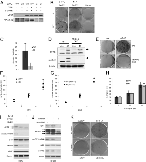
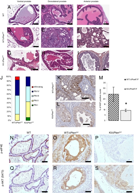
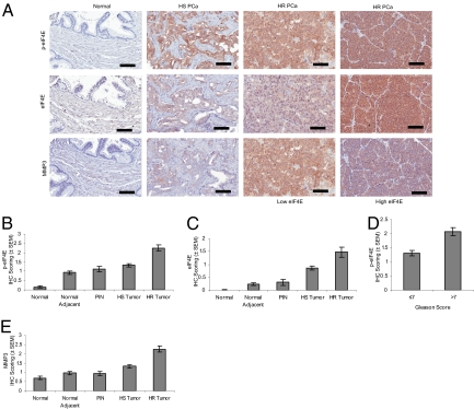
Translational regulation plays a critical role in the control of cell growth and proliferation. A key player in translational control is eIF4E, the mRNA 5' cap-binding protein. Aberrant expression of eIF4E promotes tumorigenesis and has been implicated in cancer development and progression. The activity of eIF4E is dysregulated in cancer. Regulation of eIF4E is partly achieved through phosphorylation. However, the physiological significance of eIF4E phosphorylation in mammals is not clear. Here, we show that knock-in mice expressing a nonphosphorylatable form of eIF4E are resistant to tumorigenesis in a prostate cancer model. By using a genome-wide analysis of translated mRNAs, we show that the phosphorylation of eIF4E is required for translational up-regulation of several proteins implicated in tumorigenesis. Accordingly, increased phospho-eIF4E levels correlate with disease progression in patients with prostate cancer. Our findings establish eIF4E phosphorylation as a critical event in tumorigenesis. These findings raise the possibility that chemical compounds that prevent the phosphorylation of eIF4E could act as anticancer drugs.


Conflict of interest statement

The authors declare no conflict of interest.

Figures

Fig. 1.
Fig. 1.
KI MEFs are resistant to malignant transformation. (A) Untreated or TPA-treated (100 ng/mL) MEFs were labeled with 32P-orthophosphate for 2 h as described in Materials and Methods. Cells were lysed and the supernatant was incubated with control (preimmune serum) or eIF4E antibody. Immunoprecipitated proteins were resolved by SDS/PAGE followed by autoradiography and Western blotting. (B) MEFs infected with the indicated retroviruses were grown on a monolayer and focus formation was determined after 10 d in culture by methylene blue staining. Similar results were obtained with eight independent pairs of MEFs. (C) WT and KI MEFs infected with c-MYC and RASV12 expression vectors were grown in soft agar and the total number of colonies consisting of more than eight cells were counted (six wells for each pair of MEFs). WT MEFs formed significantly more colonies than KI MEFs (two-tailed Student t test, P = 0.037). (D) Cell lysates from WT and MNK1/2 DKO MEFs expressing HA-eIF4E were resolved by SDS/PAGE followed by Western blotting. Arrows indicate the slower migrating band corresponding to the HA-tagged eIF4E. (E) MEFs infected with the indicated retroviruses were grown on a monolayer and focus formation was determined after 10 d in culture by methylene blue staining. (F and G) Cells (1 × 10⁵) were seeded on d 0 and counted on d 1, 3, and 5. Data points represent mean ± SD of three independent experiments. (H) Cells (1 × 10⁶) were seeded and subjected to treatment for 24 h with the indicated concentrations of ionomycin. Floating and attached cells were collected, and the double-positive Annexin V/PI subpopulation of cells was gated. No significant differences were observed between WT and KI MEFs (two-tailed Student t test: not treated, P = 0.511; 10 μM, P = 0.329; 15 μM, P = 0.952). Similar results were obtained in three independent experiments. (I) MEFs were serum starved for 6 h in the presence of vehicle (DMSO), PP242 (2.5 μM), or Torin1 (250 nM), serum-stimulated for 30 min, and the phosphorylation and levels of indicated proteins were determined by Western blotting. (J) Raptor was silenced in HEK293 cells by shRNA (sh-raptor). As a control, cells were infected with a lentivirus encoding a scrambled shRNA. Expression and the phosphorylation levels of the indicated proteins were determined by Western blotting. (K) WT/PTENF/F and KI/PTENF/F MEFs infected with the indicated retroviruses were grown as a monolayer and focus formation was determined after 10 d in culture by methylene blue staining. Similar results were obtained from two independent experiments each done in triplicate.
Fig. 2.
Fig. 2.
KI mice are resistant to Pten loss–induced prostate cancer. (AI) Representative H&E staining of sections from the anterior (AP), ventral (VP), and DLP lobes of the prostate of WT, WT/cPtenF/F, and KI/cPtenF/F from 5- to 6-mo-old mice (no histological difference was observed between WT and KI mice without Cre; Fig. S3). (Scale bar: 100 μm.) (J) Histograms represent percentage of predominant lesions in a cohort of seven WT/cPtenF/F and seven KI/cPtenF/F. (K and L) Representative images of Ki67 staining of DLP sections from WT/cPtenF/F and KI/cPtenF/F mice (5–6 mo old). (Scale bar: 100 μm.) (M) Slides were scanned with the Aperio Scanscope and nuclear staining was quantified with the Image Scope software nuclear algorithm. At least 4,000 nuclei per mouse were analyzed (n = 4 mice per group). Student t test (two-tailed) was performed. (NS) Ventral prostate sections from WT, WT/cPtenF/F, and KI/cPtenF/F mice were used for IHC with the indicated antibodies. (Scale bar: 50 μm.)
Fig. 3.
Fig. 3.
Differential expression of selected proteins in prostate from WT versus KI mice. (A) Serum starved WT and KI MEFs were serum-stimulated for 2 h and cell lysates were resolved by SDS/PAGE followed by Western blotting with the indicated antibodies. (B) DLP extracts were resolved by SDS/PAGE and Western blotting with the indicated antibodies was performed. (CK) Sections of the prostate from WT, WT/cPtenF/F, and KI/cPtenF/F mice were used for IHC with the indicated antibodies. VP sections were used for detection of SMA and MMP3. CCL2 was detected in the anterior prostate. (Scale bar: 50 μm.)
Fig. 4.
Fig. 4.
p-eIF4E correlates with progression to HR PCa. eIF4E, phospho-eIF4E, and MMP3 were detected by IHC in human prostate cancer TMAs. The intensity of staining was scored from 0 to 4 for each core and data were analyzed as described in Materials and Methods. (A) Representative images of staining intensity obtained for eIF4E and p-eIF4E. (Scale bar: 100 μm.) (B and C) eIF4E and phospho-eIF4E immunoreactivity shows a gradual increase in intensity from the normal, normal adjacent, PIN, HS tumor, and HR tumor tissues. Significant statistical differences were found between the different histopathological groups (P < 0.001, Kruskal-Wallis test). (D) The mean intensity difference of phospho-eIF4E in cases with Gleason score greater than 7 compared with Gleason scores of 7 or lower is statistically significant (P < 0.05, Mann–Whitney U test). (E) MMP3 immunoreactivity is gradually increased in intensity from PIN to HS tumor and HR tumor tissues. Significant statistical differences exist between the different histopathological groups (P < 0.001, Kruskal-Wallis test).

Comment in

Similar articles

Cited by

References

    1. Mamane Y, Petroulakis E, LeBacquer O, Sonenberg N. mTOR, translation initiation and cancer. Oncogene. 2006;25:6416–6422. - PubMed
    1. De Benedetti A, Graff JR. eIF-4E expression and its role in malignancies and metastases. Oncogene. 2004;23:3189–3199. - PubMed
    1. Clemens MJ. Targets and mechanisms for the regulation of translation in malignant transformation. Oncogene. 2004;23:3180–3188. - PubMed
    1. Schneider RJ, Sonenberg N. Translational control in cancer development. In: Mathews MB, Sonenberg N, Hershey JWB, editors. Control in Biology and Medicine. Cold Spring Harbor, NY: Cold Spring Harbor Lab Press; 2007. pp. 401–432.
    1. Lazaris-Karatzas A, Montine KS, Sonenberg N. Malignant transformation by a eukaryotic initiation factor subunit that binds to mRNA 5′ cap. Nature. 1990;345:544–547. - PubMed

Publication types

Substances

Associated data