

A tumor-suppressing mechanism in Drosophila involving cell competition and the Hippo pathway
- PMID: 20679206
- PMCID: PMC2930466
- DOI: 10.1073/pnas.1009376107
A tumor-suppressing mechanism in Drosophila involving cell competition and the Hippo pathway
Abstract
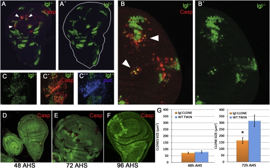
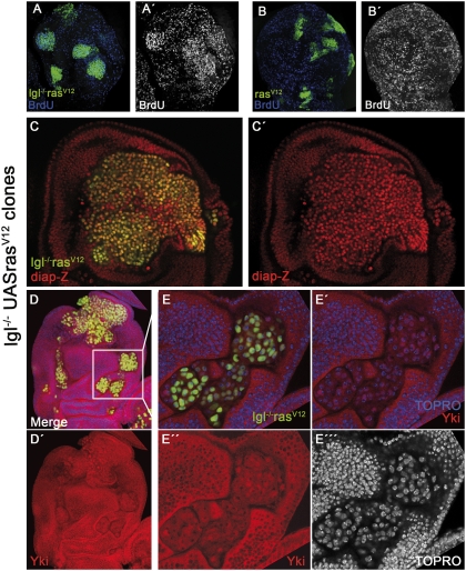
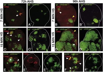
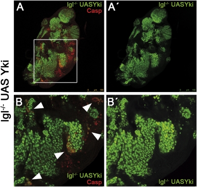
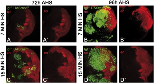
Mutant larvae for the Drosophila gene lethal giant larva (lgl) develop neoplastic tumors in imaginal discs. However, lgl mutant clones do not form tumors when surrounded by wild-type tissue, suggesting the existence of a tumor-suppressing mechanism. We have investigated the tumorigenic potential of lgl mutant cells by generating wing compartments that are entirely mutant for lgl and also inducing clones of various genetic combinations of lgl(-) cells. We find that lgl(-) compartments can grow indefinitely but lgl(-) clones are eliminated by cell competition. lgl mutant cells may form tumors if they acquire constitutive activity of the Ras pathway (lgl(-) UAS-ras(V12)), which confers proliferation advantage through inhibition of the Hippo pathway. Yet, the majority of lgl(-) UAS-ras(V12) clones are eliminated in spite of their high proliferation rate. The formation of a tumor requires in addition the formation of a microenvironment that allows mutant cells to evade cell competition.
Conflict of interest statement
The authors declare no conflict of interest.
Figures
Comment in
-
Tumorigenesis: To the death!Nat Rev Cancer. 2010 Sep;10(9):598. doi: 10.1038/nrc2924. Nat Rev Cancer. 2010. PMID: 20803809 No abstract available.
Similar articles
-
Epithelial neoplasia in Drosophila entails switch to primitive cell states.Proc Natl Acad Sci U S A. 2013 Jun 11;110(24):E2163-72. doi: 10.1073/pnas.1212513110. Epub 2013 May 24. Proc Natl Acad Sci U S A. 2013. PMID: 23708122 Free PMC article.
-
Loss of the Drosophila cell polarity regulator Scribbled promotes epithelial tissue overgrowth and cooperation with oncogenic Ras-Raf through impaired Hippo pathway signaling.BMC Dev Biol. 2011 Sep 29;11:57. doi: 10.1186/1471-213X-11-57. BMC Dev Biol. 2011. PMID: 21955824 Free PMC article.
-
Lgl, aPKC, and Crumbs regulate the Salvador/Warts/Hippo pathway through two distinct mechanisms.Curr Biol. 2010 Apr 13;20(7):573-81. doi: 10.1016/j.cub.2010.01.055. Epub 2010 Apr 1. Curr Biol. 2010. PMID: 20362447
-
Lgl/aPKC and Crb regulate the Salvador/Warts/Hippo pathway.Fly (Austin). 2010 Oct-Dec;4(4):288-93. doi: 10.4161/fly.4.4.13116. Epub 2010 Oct 21. Fly (Austin). 2010. PMID: 20798605 Free PMC article. Review.
-
Deciphering tumor-suppressor signaling in flies: genetic link between Scribble/Dlg/Lgl and the Hippo pathways.J Genet Genomics. 2011 Oct 20;38(10):461-70. doi: 10.1016/j.jgg.2011.09.005. Epub 2011 Sep 17. J Genet Genomics. 2011. PMID: 22035867 Review.
Cited by
-
Aerobic glycolysis tunes YAP/TAZ transcriptional activity.EMBO J. 2015 May 12;34(10):1349-70. doi: 10.15252/embj.201490379. Epub 2015 Mar 21. EMBO J. 2015. PMID: 25796446 Free PMC article.
-
Tumorigenesis: To the death!Nat Rev Cancer. 2010 Sep;10(9):598. doi: 10.1038/nrc2924. Nat Rev Cancer. 2010. PMID: 20803809 No abstract available.
-
Apical deficiency triggers JNK-dependent apoptosis in the embryonic epidermis of Drosophila.Development. 2011 Jul;138(14):3021-31. doi: 10.1242/dev.059980. Development. 2011. PMID: 21693518 Free PMC article.
-
Systemic organ wasting induced by localized expression of the secreted insulin/IGF antagonist ImpL2.Dev Cell. 2015 Apr 6;33(1):36-46. doi: 10.1016/j.devcel.2015.02.012. Dev Cell. 2015. PMID: 25850671 Free PMC article.
-
Mutant p53 in cell-cell interactions.Genes Dev. 2021 Apr 1;35(7-8):433-448. doi: 10.1101/gad.347542.120. Genes Dev. 2021. PMID: 33861719 Free PMC article. Review.
References
-
- Bilder D. Epithelial polarity and proliferation control: Links from the Drosophila neoplastic tumor suppressors. Genes Dev. 2004;18:1909–1925. - PubMed
-
- Hariharan IK, Bilder D. Regulation of imaginal disc growth by tumor-suppressor genes in Drosophila. Annu Rev Genet. 2006;40:335–361. - PubMed
-
- Knoblich JA. Mechanisms of asymmetric stem cell division. Cell. 2008;132:583–597. - PubMed
-
- Humbert PO, et al. Control of tumourigenesis by the Scribble/Dlg/Lgl polarity module. Oncogene. 2008;27:6888–6907. - PubMed
-
- Kuphal S, et al. Expression of Hugl-1 is strongly reduced in malignant melanoma. Oncogene. 2006;25:103–110. - PubMed
Publication types
MeSH terms
Substances
LinkOut - more resources
Full Text Sources
Molecular Biology Databases
