. 2010 Aug 17;107(33):14775-80.
doi: 10.1073/pnas.0914206107. Epub 2010 Aug 2.

A canine Arylsulfatase G (ARSG) mutation leading to a sulfatase deficiency is associated with neuronal ceroid lipofuscinosis

Affiliations

A canine Arylsulfatase G (ARSG) mutation leading to a sulfatase deficiency is associated with neuronal ceroid lipofuscinosis

Marie Abitbol et al. Proc Natl Acad Sci U S A. .

Abstract

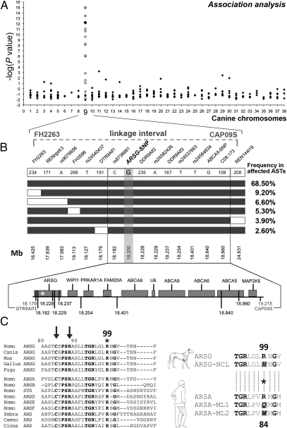
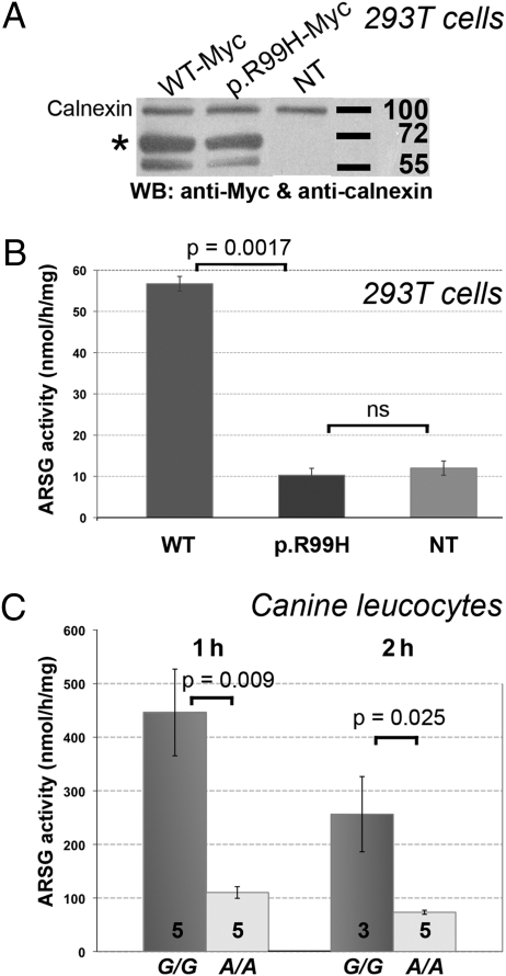
Neuronal ceroid lipofuscinoses (NCLs) represent the most common group of inherited progressive encephalopathies in children. They are characterized by progressive loss of vision, mental and motor deterioration, epileptic seizures, and premature death. Rare adult forms of NCL with late onset are known as Kufs' disease. Loci underlying these adult forms remain unknown due to the small number of patients and genetic heterogeneity. Here we confirm that a late-onset form of NCL recessively segregates in US and French pedigrees of American Staffordshire Terrier (AST) dogs. Through combined association, linkage, and haplotype analyses, we mapped the disease locus to a single region of canine chromosome 9. We eventually identified a worldwide breed-specific variant in exon 2 of the Arylsulfatase G (ARSG) gene, which causes a p.R99H substitution in the vicinity of the catalytic domain of the enzyme. In transfected cells or leukocytes from affected dogs, the missense change leads to a 75% decrease in sulfatase activity, providing a functional confirmation that the variant might be the NCL-causing mutation. Our results uncover a protein involved in neuronal homeostasis, identify a family of candidate genes to be screened in patients with Kufs' disease, and suggest that a deficiency in sulfatase is part of the NCL pathogenesis.


Conflict of interest statement

Conflict of interest statement: INRA and ENVA have applied for a patent covering the use of the canine ARSG SNP for the diagnosis of NCL or for selective breeding of dogs. M.A. and S. Blot are listed as inventors in this application.

Figures

Fig. 1.
Fig. 1.
Clinical and histopathological features of the disease. (A) The wide-based stance (white polygon) of a 5-y-old affected AST illustrates loss of motor coordination. (B) Sagittal 2-T weighted MRI of the brain from a 6-y-old AST through the cerebellum. (Inset) A similar image at the same scale of the cerebellum from an age-matched healthy AST, the outline of which has been projected on the cerebellum of the main image (white dotted line). A reduction of gray matter is demonstrated by the enlarged sulci (arrowheads). (C and D) Transverse sections of the cerebellum from healthy dogs (C) and affected dogs (D), stained with PAS reagent and counterstained in Mayer's hematoxylin solution. (C) In a normal cerebellum, the large PAS-negative Purkinje neurons lie between the molecular (top left) and granular (bottom right) layers. (D) In the cerebellum from an affected dog, massive Purkinje cell loss results in a blurry line. The remaining Purkinje neurons (arrows) or neuron remnants (arrowheads) accumulate perinuclear PAS-positive granular material. (Inset) Affected Purkinje neurons imaged on transmission electron microscopy show accumulated lysosomal material composed of concentric straight or curved profiles with alternating clear and dense bands. (Scale bars: 50 μm for the sections; 250 nm for the inset.)
Fig. 2.
Fig. 2.
Mapping, fine-mapping, and identification of a candidate gene for NCL in ASTs. (A) A single locus with strong genome-wide significance was identified on CFA09 (larger black diamonds) and confirmed (white diamonds); see Table S2. The −log(P values) are reported on the y-axis. The 2.8-Mb candidate region defined by our linkage analysis (18) is shown. (B) Fine-mapping using haplotype analysis for NCL. Haplotypes identified in affected ASTs are shown as boxes (NCL allele, black boxes; alternative allele, white boxes). Genotyped microsatellites and SNPs are indicated in their 5′-3′ position, and their position on CFA09 is shown. The Haplotypes with a frequency <2% are omitted. In the bottom part, candidate genes within the critical region are shown. The relative positions of markers are indicated by vertical black lines, and the nonsynonymous associated ARSG-SNP is represented by an asterisk. (C) (Left) N-terminal sequence of ARSG or predicted ARS from metazoans is aligned with the N-terminal amino acids of human ARS. Conserved amino acids are in bold type. Arrows point to two of the 10 residues involved in the catalytic site of arylsulfatases (22), with positions referring to the canine ARSG. The R99 mutant in affected dogs is highlighted by an asterisk. (Right) The sequence flanking the R99 of the canine WT ARSG protein (ARSG) is aligned with the corresponding sequence of the mutated protein (ARSG-NCL) and best aligned with the R84 of the human WT ARSA (ARSA), replaced by a glutamine (Q) or a tryptophane (W) in some patients affected by metachromatic leukodystrophy [ARSA-ML1; (25) and ARSA-ML2, (24)].
Fig. 3.
Fig. 3.
The highly conserved R99 is critical for ARSG activity. (A) In HEK293T cells, similar overexpression levels of C-ter Myc-tagged WT (WT-Myc) or variant (p.R99H-Myc) human ARSG are validated in a Western blot assay (WB) by their specific immunoreactivity with an anti-Myc antibody (asterisk). Proteins are detected at the expected molecular weight (63 kDa). NT, nontransfected cells. Adjusted levels of total proteins are confirmed by levels of the housekeeping calnexin. (B) These HEK293T cells were used to measure ARSG activity, expressed in nmol/h/mg of protein, using a method adapted from Frese et al. (32). Vertical bars represent the SEM for seven experiments. (C) Total leukocyte arylsulfatase activity was compared between homozygous (G/G) healthy controls, taken as the 100% reference activity level, and homozygous (A/A) affected ASTs. The number of sampled dogs is identified within each histogram, and vertical bars represent SEM.

Similar articles

Cited by

References

    1. Jalanko A, Braulke T. Neuronal ceroid lipofuscinoses. Biochim Biophys Acta. 2009;1793:697–709. - PubMed
    1. Berkovic SF, Carpenter S, Andermann F, Andermann E, Wolfe LS. Kufs’’ disease: A critical reappraisal. Brain. 1988;111:27–62. - PubMed
    1. Haltia M. The neuronal ceroid-lipofuscinoses: From past to present. Biochim Biophys Acta. 2006;1762:850–856. - PubMed
    1. Lewandowska E, et al. Kufs’’ disease: Diagnostic difficulties in the examination of extracerebral biopsies. Folia Neuropathol. 2009;47:259–267. - PubMed
    1. Bellettato CM, Scarpa M. Pathophysiology of neuropathic lysosomal storage disorders. J Inherit Metab Dis. 2010;33:347–362. - PubMed

Publication types

MeSH terms

Substances

LinkOut - more resources