

Cooperation of Tim-3 and PD-1 in CD8 T-cell exhaustion during chronic viral infection
- PMID: 20679213
- PMCID: PMC2930455
- DOI: 10.1073/pnas.1009731107
Cooperation of Tim-3 and PD-1 in CD8 T-cell exhaustion during chronic viral infection
Abstract
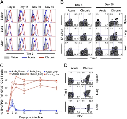
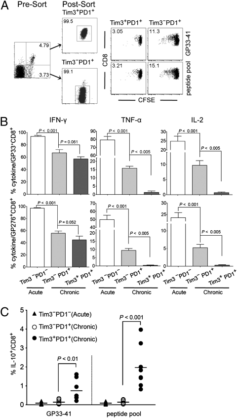
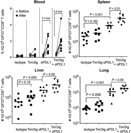
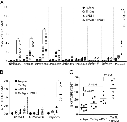
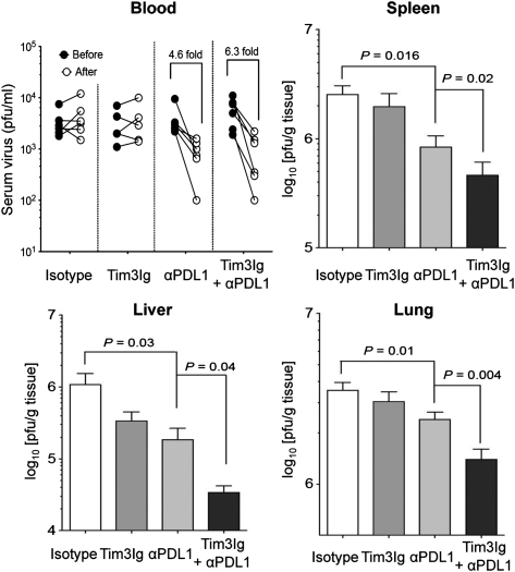
Inhibitory receptors play a crucial role in regulating CD8 T-cell function during chronic viral infection. T-cell Ig- and mucin-domain-containing molecule-3 (Tim-3) is well known to negatively regulate T-cell responses, but its role in CD8 T-cell exhaustion during chronic infection in vivo remains unclear. In this study, we document coregulation of CD8 T cell exhaustion by Tim-3 and PD-1 during chronic lymphocytic choriomeningitis virus infection. Whereas Tim-3 was only transiently expressed by CD8 T cells after acute infection, virus-specific CD8 T cells retained high Tim-3 expression throughout chronic infection. The majority (approximately 65% to 80%) of lymphocytic choriomeningitis virus-specific CD8 T cells in lymphoid and nonlymphoid organs coexpressed Tim-3 and PD-1. This coexpression of Tim-3 and PD-1 was associated with more severe CD8 T-cell exhaustion in terms of proliferation and secretion of effector cytokines such as IFN-gamma, TNF-alpha, and IL-2. Interestingly, CD8 T cells expressing both inhibitory receptors also produced the suppressive cytokine IL-10. Most importantly, combined blockade of Tim-3 and PD-1 pathways in vivo synergistically improved CD8 T cell responses and viral control in chronically infected mice. Taken together, our study defines a parameter for determining the severity of CD8 T cell dysfunction and for identifying virus-specific CD8 T cells that produce IL-10, and shows that targeting both PD-1 and Tim-3 is an effective immune strategy for treating chronic viral infections.
Conflict of interest statement
Conflict of interest statement: R.A. received licensing fees from Genentech on using antibodies to block the PD-1 inhibitory pathway. G.J.F. received patent royalties on the PD-1 and TIM-3 pathways. No other authors have any financial conflicts.
Figures
References
-
- Shin H, Wherry EJ. CD8 T cell dysfunction during chronic viral infection. Curr Opin Immunol. 2007;19:408–415. - PubMed
-
- Barber DL, et al. Restoring function in exhausted CD8 T cells during chronic viral infection. Nature. 2006;439:682–687. - PubMed
-
- Sharpe AH, Wherry EJ, Ahmed R, Freeman GJ. The function of programmed cell death 1 and its ligands in regulating autoimmunity and infection. Nat Immunol. 2007;8:239–245. - PubMed
Publication types
MeSH terms
Substances
Grants and funding
- P01 AI056299/AI/NIAID NIH HHS/United States
- AI73748/AI/NIAID NIH HHS/United States
- AI08080192/AI/NIAID NIH HHS/United States
- P01 AI073748/AI/NIAID NIH HHS/United States
- R01 AI089955/AI/NIAID NIH HHS/United States
- R29 NS030843/NS/NINDS NIH HHS/United States
- P01 NS038037/NS/NINDS NIH HHS/United States
- R01 NS030843/NS/NINDS NIH HHS/United States
- NS030843/NS/NINDS NIH HHS/United States
- R01 NS045937/NS/NINDS NIH HHS/United States
- NS054096/NS/NINDS NIH HHS/United States
- K01 NS054096/NS/NINDS NIH HHS/United States
- NS038037/NS/NINDS NIH HHS/United States
- P01 AI054456/AI/NIAID NIH HHS/United States
- R01 AI139675/AI/NIAID NIH HHS/United States
- R37 NS030843/NS/NINDS NIH HHS/United States
- AI054456/AI/NIAID NIH HHS/United States
- NS045937/NS/NINDS NIH HHS/United States
- AI056299/AI/NIAID NIH HHS/United States
LinkOut - more resources
Full Text Sources
Other Literature Sources
Molecular Biology Databases
Research Materials
