

A mammalian neural tissue opsin (Opsin 5) is a deep brain photoreceptor in birds
- PMID: 20679218
- PMCID: PMC2930557
- DOI: 10.1073/pnas.1006393107
A mammalian neural tissue opsin (Opsin 5) is a deep brain photoreceptor in birds
Abstract
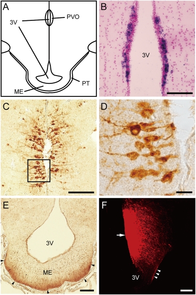
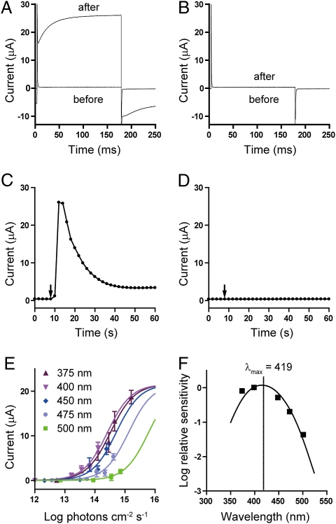
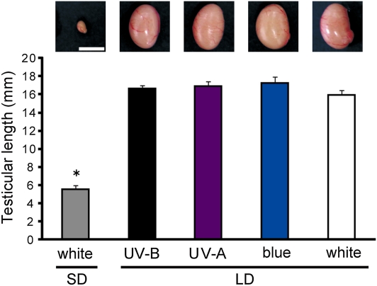
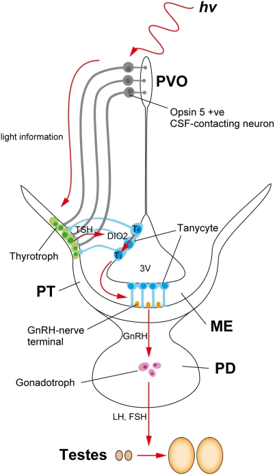
It has been known for many decades that nonmammalian vertebrates detect light by deep brain photoreceptors that lie outside the retina and pineal organ to regulate seasonal cycle of reproduction. However, the identity of these photoreceptors has so far remained unclear. Here we report that Opsin 5 is a deep brain photoreceptive molecule in the quail brain. Expression analysis of members of the opsin superfamily identified as Opsin 5 (OPN5; also known as Gpr136, Neuropsin, PGR12, and TMEM13) mRNA in the paraventricular organ (PVO), an area long believed to be capable of phototransduction. Immunohistochemistry identified Opsin 5 in neurons that contact the cerebrospinal fluid in the PVO, as well as fibers extending to the external zone of the median eminence adjacent to the pars tuberalis of the pituitary gland, which translates photoperiodic information into neuroendocrine responses. Heterologous expression of Opsin 5 in Xenopus oocytes resulted in light-dependent activation of membrane currents, the action spectrum of which showed peak sensitivity (lambda(max)) at approximately 420 nm. We also found that short-wavelength light, i.e., between UV-B and blue light, induced photoperiodic responses in eye-patched, pinealectomized quail. Thus, Opsin 5 appears to be one of the deep brain photoreceptive molecules that regulates seasonal reproduction in birds.
Conflict of interest statement
The authors declare no conflict of interest.
Figures
Comment in
-
Shedding light on photoperiodism.Proc Natl Acad Sci U S A. 2010 Sep 7;107(36):15662-3. doi: 10.1073/pnas.1010370107. Epub 2010 Aug 27. Proc Natl Acad Sci U S A. 2010. PMID: 20802157 Free PMC article. No abstract available.
References
-
- Oliver J, Bayle JD. Brain photoreceptors for the photoinduced testicular response in birds. Experientia. 1982;38:1020–1029. - PubMed
-
- von Frisch K. Beitrage zur physiologie der pigmentzellen in der fischhaut. Pflugers Arch Gesamte Physiol Menschen Tiere. 1911;138:319–387.
-
- Benoit J. Le role des yeux dans l'action stimulante de la lumiere sure le developpement testiulaire chez le canard. CR Soc Biol (Paris) 1935;118:669–671.
-
- Follett BK, King VM, Meddle SL. In: Biological Rhythms and Photoperiodism in Plants. Lumsden PJ, Miller AJ, editors. Oxford: BIOS; 1998. pp. 231–242.
Publication types
MeSH terms
Substances
Associated data
- Actions
LinkOut - more resources
Full Text Sources
Other Literature Sources
Molecular Biology Databases
Miscellaneous
