. 2010 Aug 17;107(33):14745-50.
doi: 10.1073/pnas.1001562107. Epub 2010 Aug 2.

CX3CR1+ CD8alpha+ dendritic cells are a steady-state population related to plasmacytoid dendritic cells

Affiliations

CX3CR1+ CD8alpha+ dendritic cells are a steady-state population related to plasmacytoid dendritic cells

Liat Bar-On et al. Proc Natl Acad Sci U S A. .

Abstract

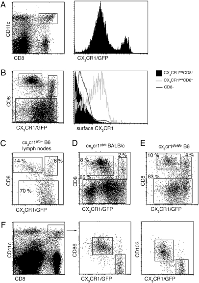
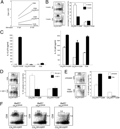
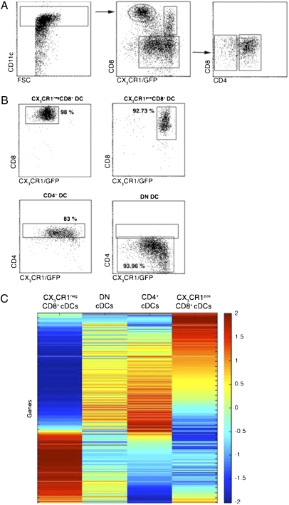
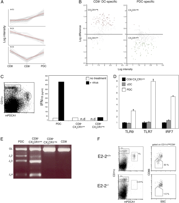
Lymphoid organs are characterized by a complex network of phenotypically distinct dendritic cells (DC) with potentially unique roles in pathogen recognition and immunostimulation. Classical DC (cDC) include two major subsets distinguished in the mouse by the expression of CD8alpha. Here we describe a subset of CD8alpha(+) DC in lymphoid organs of naïve mice characterized by expression of the CX(3)CR1 chemokine receptor. CX(3)CR1(+) CD8alpha(+) DC lack hallmarks of classical CD8alpha(+) DC, including IL-12 secretion, the capacity to cross-present antigen, and their developmental dependence on the transcriptional factor BatF3. Gene-expression profiling showed that CX(3)CR1(+) CD8alpha(+) DC resemble CD8alpha(-) cDC. The microarray analysis further revealed a unique plasmacytoid DC (PDC) gene signature of CX(3)CR1(+) CD8alpha(+) DC. A PDC relationship of the cells is supported further by the fact that they harbor characteristic D-J Ig gene rearrangements and that development of CX(3)CR1(+) CD8alpha(+) DC requires E2-2, the critical transcriptional regulator of PDC. Thus, CX(3)CR1(+) CD8alpha(+) DC represent a unique DC subset, related to but distinct from PDC. Collectively, the expression-profiling data of this study refine the resolution of previous DC definitions, sharpen the border of classical CD8alpha(+) and CD8alpha(-) DC, and should assist the identification of human counterparts of murine DC subsets.


Conflict of interest statement

The authors declare no conflict of interest.

Figures

Fig. 1.
Fig. 1.
Identification of the CX3CR1+ CD8α+ DC subset. (A) Flow cytometric analysis of splenocytes from Cx3cr1gfp/+ mice. cDC were identified as CD11chi cells. Histogram shows GFP expression in CD11chi CD8α+ DC. (B) Staining of splenic cDC subpopulations for surface expression of CX3CR1 using a CX3CL1-Fc fusion protein. (C) Flow cytometric analysis of lymph node cells from Cx3cr1gfp/+ mouse. (D) Flow cytometric analysis of splenocytes from Cx3cr1gfp/+ BALB/c mouse. (E) Flow cytometric analysis of splenocytes from CX3CR1-deficient Cx3cr1gfp/gfp mice. (F) Flow cytometric analysis of splenocytes from Cx3cr1gfp/+ mouse. Note characterization of CX3CR1/GFP+ CD8α+ DC as a CD86lo CD103neg population.
Fig. 2.
Fig. 2.
CX3CR1+ CD8α+ DC lack hallmarks of classical CD8α+ DC. (A) Mixed leukocyte reaction with indicated numbers of sorted splenic DC subsets isolated from Cx3cr1gfp/+ C57BL/6 mice and BALB/c CD4+ T cells (105). Data are representative of two experiments. (B) Flow cytometric analysis of splenic DC subsets in 3- and 8-wk-old Cx3cr1gfp/+ C57BL/6 mice. Bars represent the percentages of CX3CR1 CD8α+ and CX3CR1+ CD8α+ subsets out of total cDC (CD11chi cells). Data are representative of two experiments. (C) Analysis of IL-12p70 (Left) and IL-12p40 (Right) secretion by sorted splenic DC subsets in response to in vitro exposure to CpG. Data are representative of two experiments.(D) Selective deletion of CX3CR1 CD8α+ splenic DC by CytC injection. Bars represent the percentages of CX3CR1 CD8α+ and CX3CR1+ CD8α+ subsets out of total DC (CD11chi cells). Data are representative of two experiments. (E) Flow cytometric analysis of splenic DC subsets of Cx3cr1gfp/+ C57BL/6 mice bearing a WT tumor or a tumor secreting FLt3L. Bars represent the percentages of CX3CR1 CD8α+ and CX3CR1+ CD8α+ subsets out of total cDC (CD11chi cells) (n = 3). Data are representative of two experiments. (F) Flow cytometric analysis of splenic DC from Batf3+/+Cx3cr1gfp/gfp, Batf3+/−Cx3cr1gfp/gfp, and Batf3−/−Cx3cr1gfp/gfp mice. cDC were gated as CD11chi B220 cells.
Fig. 3.
Fig. 3.
Sorting strategy and gene-expression matrix of splenic cDC subsets. (A) Flow cytometric analysis of magnetic bead-enriched CD11chi cells isolated from Cx3cr1gfp/+ mice indicating sorting gates. (B) Analysis of sorted DC subsets. Percentages indicate purity of the respective cDC populations. (C) Expression matrix of the 500 modulated genes. Rows represent individual genes, and columns represent splenic cDC subsets. Colors indicate the relative expression levels of the genes in the different subsets, according to the code shown on the right.
Fig. 4.
Fig. 4.
CX3CR1+ CD8α+ DC share expression profile, somatic Ig gene rearrangements, and E2-2 dependence with PDC. (A) PCA of the genes overexpressed in CX3CR1+ CD8α+ cells compared with CD8α+ cDC. Shown are the three principal component sets of genes analyzed against the expression data of Robbins et al. (35). Note the preferential expression in PDC (Top), in CD8α DC (Middle), and in both PDC and CD8α DC (Bottom). (B) Distribution of genes specific for CD8α+ DC or PDC in the two CD8α+ DC subsets. Shown is pairwise comparison of average probe intensities in CX3CR1+ and CX3CR1 CD8α+ DC; genes specific for CD8α+ DC or PDC were selected based on reference . (C) IFN-α production by sorted splenic DC subsets after incubation for 20 h in the presence or absence of 400 HAU/mL influenza virus. (D) Expression of the TLR/IFN signaling genes in sorted CD8α cDC, PDC, and CX3CR1+ CD8α+ DC as determined by quantitative RT-PCR (mean ± SD of triplicate reactions). Expression levels are normalized relative to the CX3CR1+CD8α+ subset. (E) D–J rearrangement of IgH gene in sorted DC subsets. IgH D–J rearrangements were detected by genomic PCR using primer sets for the DHQ52 element. Data are representative of three experiments. (F) Absence of CX3CR1+ CD8α+ DC and PDC in absence of the transcription factor E2-2. (Left) Gated donor-derived (CD45.2+) splenocytes from E2-2−/− chimeras or control E2-2+/+ chimeras mice were analyzed for the presence of CD11cint Bst2+ PDC. (Right) Gated CD11chi CD8α+ DC comprise the CD86lo SSClo and the CD86hi SSChi subsets (corresponding to CX3CR1+ and CX3CR1 populations, respectively). Data are representative of three independent experiments.

Similar articles

Cited by

References

    1. Sapoznikov A, Jung S. Probing in vivo dendritic cell functions by conditional cell ablation. Immunol Cell Biol. 2008;86:409–415. - PubMed
    1. Steinman RM, Witmer MD. Lymphoid dendritic cells are potent stimulators of the primary mixed leukocyte reaction in mice. Proc Natl Acad Sci USA. 1978;75:5132–5136. - PMC - PubMed
    1. Liu K, et al. In vivo analysis of dendritic cell development and homeostasis. Science. 2009;324:392–397. - PMC - PubMed
    1. Shortman K, Naik SH. Steady-state and inflammatory dendritic-cell development. Nat Rev Immunol. 2007;7:19–30. - PubMed
    1. Dudziak D, et al. Differential antigen processing by dendritic cell subsets in vivo. Science. 2007;315:107–111. - PubMed

Publication types

MeSH terms

Associated data