. 2010 Aug 17;107(33):14805-10.
doi: 10.1073/pnas.1002119107. Epub 2010 Aug 2.

Intermolecular associations determine the dynamics of the circadian KaiABC oscillator

Affiliations

Intermolecular associations determine the dynamics of the circadian KaiABC oscillator

Ximing Qin et al. Proc Natl Acad Sci U S A. .

Abstract

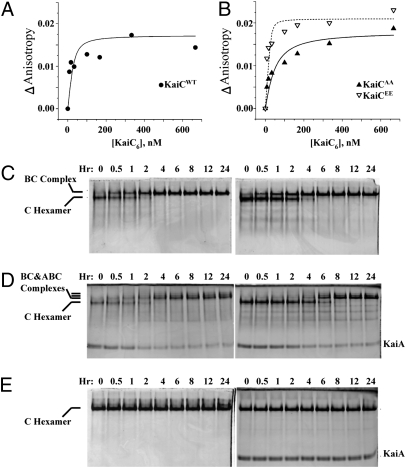
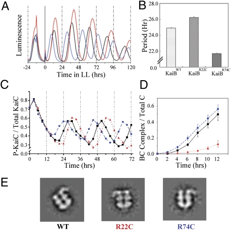
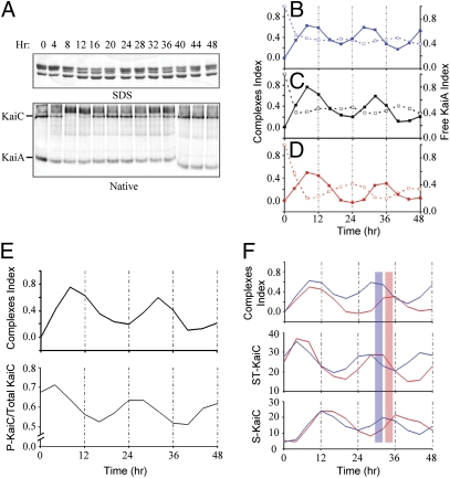
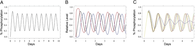
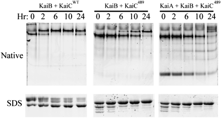
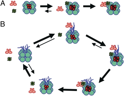
Three proteins from cyanobacteria (KaiA, KaiB, and KaiC) can reconstitute circadian oscillations in vitro. At least three molecular properties oscillate during this reaction, namely rhythmic phosphorylation of KaiC, ATP hydrolytic activity of KaiC, and assembly/disassembly of intermolecular complexes among KaiA, KaiB, and KaiC. We found that the intermolecular associations determine key dynamic properties of this in vitro oscillator. For example, mutations within KaiB that alter the rates of binding of KaiB to KaiC also predictably modulate the period of the oscillator. Moreover, we show that KaiA can bind stably to complexes of KaiB and hyperphosphorylated KaiC. Modeling simulations indicate that the function of this binding of KaiA to the KaiB*KaiC complex is to inactivate KaiA's activity, thereby promoting the dephosphorylation phase of the reaction. Therefore, we report here dynamics of interaction of KaiA and KaiB with KaiC that determine the period and amplitude of this in vitro oscillator.


Conflict of interest statement

The authors declare no conflict of interest.

Figures

Fig. 1.
Fig. 1.
Interactions among KaiA, KaiB, and KaiC. (A and B) Fluorescence anisotropy was used to calculate the binding affinity between KaiA and KaiC. (A) FA between KaiA and KaiCWT. F150-labeled KaiA (60 nM) was mixed with increasing concentrations of unlabeled KaiCWT proteins, and the anisotropy was measured. The dissociation constant (Kd) was calculated by assuming a 1:1 ratio binding stoichiometry between KaiA dimers and KaiC hexamers by the equation in ref. and simulated by the curves shown on the panels. (B) FA between KaiA (60 nM) and KaiCAA or KaiCEE phosphomimics. (C–E) Formation of stable complexes among KaiA, KaiB, and KaiC was assayed by native PAGE. (C) Formation of stable complexes of KaiB with KaiCDT (Left) or KaiCEE (Right), as indicated by a reduction in the mobility of the KaiC band. (D) In the presence of KaiB, KaiA can be included in a stable KaiA•KaiB•KaiC complex, as indicated by extra bands and the depletion of the free KaiA dimer band (KaiCDT, Left, and KaiCEE, Right). (E) In the case of a KaiC variant that cannot be phosphorylated on the S431 residue (KaiCAT), stable KaiB•KaiCAT (Left) or KaiA•KaiB•KaiCAT complexes do not form.
Fig. 2.
Fig. 2.
KaiB mutant variants that affect association kinetics also affect circadian period in vivo and in vitro. In this figure, black traces are KaiBWT, red traces are KaiBR22C (long period), and blue traces are KaiBR74C (short period). (A) Luminescence traces of cyanobacterial strains expressing different KaiB variant proteins in vivo. (B) Quantitative analysis of the periods in A: KaiBWT = 24.9 ± 0.10 h; KaiBR22C = 26.2 ± 0.12 h; KaiBR74C = 21.7 ± 0.10 h (mean ± SD, n = 3 for each variant). (C) In vitro rhythms with KaiAWT + KaiCWT and each of the three KaiB mutant variants. The mutations of KaiB show similar effects on the in vitro rhythm of KaiC phosphorylation as on the in vivo rhythm of gene expression as reported by luminescence in A. (D) Formation of complexes between KaiC489 and the three KaiB mutant variants (±SD, n = 3; for raw data, see SI Appendix, Fig. S3). (E) Electron microscopy analyses of the stable KaiA•KaiB•KaiC complexes show similar configurations among the different KaiB variants.
Fig. 3.
Fig. 3.
Rhythmic assembly of KaiA•KaiB•KaiC complexes quantitatively correlate with KaiC phosphostatus. (A, Upper) Using KaiAWT, KaiBWT, and KaiCWT, samples were collected every 4 h during the in vitro oscillation and analyzed by SDS/PAGE and native PAGE. Each lane of the SDS/PAGE gel clearly shows four bands representing the four phosphoforms of KaiCWT (19, 20). These data are quantified in E (Lower). (A, Lower) The same samples were run on native PAGE, and low-mobility KaiA•KaiB•KaiC complexes appear and disappear rhythmically in antiphase to the density of the KaiC hexamer and KaiA dimer bands (the image comes from two separate gels, hence the discontinuity of the last three lanes). (B) Quantification of the formation of complexes (solid line) and free KaiA (dashed line) in the native PAGE for KaiBR74C (raw data appears in SI Appendix, Fig. S4A). (C) Same as for B except using KaiBWT (raw data appears in A). (D) Same as for B except using KaiBR22C (raw data appears in SI Appendix, Fig. S4B). The period of the rhythm of Kai protein complex formation is comparable to the in vivo luminescence rhythm and the in vitro KaiC phosphorylation rhythms. (E) The formation of complexes (Upper) is compared with the KaiC phosphorylation rhythm (Lower) (raw data in A). Formation of KaiA•KaiB•KaiC complexes peaks in the phase of KaiC dephosphorylation. (F) Relationship between formation of complexes and different KaiC phosphoforms. The analysis of KaiC phosphorylation status and phosphoforms was derived from SDS/PAGE gels as shown in SI Appendix, Figs. S1 and S5). The short period mutant KaiBR74C is plotted with the long period mutant KaiBR22C to show the maximum contrast (the peaks for KaiBWT were intermediate between those for KaiBR74C and KaiBR22C, as shown in SI Appendix, Fig. S5). The formation of KaiA•KaiB•KaiC complexes (Top), doubly phosphorylated KaiC (S431-P and T432-P; Middle), and singly phosphorylated KaiC (S431-P; Bottom) were plotted as a function of incubation time. The blue bar is a reference for the peak of KaiA•KaiBR74C•KaiC complex formation, whereas the red bar is a reference for the peak of KaiA•KaiBR22C•KaiC complex formation.
Fig. 4.
Fig. 4.
Computational model of the dynamics of KaiABC complexes. (A) Typical simulation from the simplified mathematical model of the net KaiC population phosphorylation with standard initial conditions of [KaiA dimer]:[KaiB tetramer]:[KaiC hexamer] = 1:1.5:1 and setting C0 = 1 μM. (B) Kinetics of sequestration of KaiA in high-molecular-weight complexes (with KaiB and KaiC) shows the rapid formation of labile KaiA•KaiC complexes (red trace) followed by sequestration into stable KaiA•KaiB•KaiC complexes (blue trace). The black trace is net KaiC phosphorylation. (C) The simulated effect of varying the association kinetics of KaiB to hyperphosphorylated KaiC on net KaiC phosphorylation shows a correlation between an increased rate of association (5.0 = green, 2.5 = red, 1.0 = black, 0.5 = yellow, 0.25 = blue) and a shorter circadian period.
Fig. 5.
Fig. 5.
KaiC489 forms stable complexes with KaiB and KaiA. KaiC489 (from which the 30 amino acids that form “tentacles” at the C terminus of KaiC were deleted) forms a hexamer that can assemble into a stable KaiB•KaiC complex (Center) as well as a stable KaiA•KaiB•KaiC complex (Right). KaiCWT also forms stable KaiB•KaiC complexes (Left). These data indicate that the formation of the stable complex of KaiA with KaiC in the presence of KaiB is independent of KaiC's C-terminal tentacles. Note from the SDS/PAGE analyses at the bottom of the figure that KaiCWT dephosphorylates over time at 30 °C (Left), but KaiC489 hyperphosphorylates over time at 30 °C (Center) independently of the presence of KaiA.
Fig. 6.
Fig. 6.
KaiB•KaiC sequesters KaiA. (A) KaiC489 (“double-donut” hexamer) cannot associate with KaiA (red dimers) by itself, but once it has become hyperphosphorylated (phosphorylated residues indicated by red dots within the KaiC hexamer), it can form stable complexes with KaiB (tetramer of green diamonds) that are then able to recruit KaiA into KaiA•KaiB•KaiC complexes. (B) With KaiCWT (“double-donut” hexamer with C-terminal “tentacles”) in the cycling reaction, KaiA repeatedly and rapidly interacts with KaiC's C-terminal tentacles during the phosphorylation phase. When KaiCWT becomes hyperphosphorylated, it first binds KaiB stably. Then the KaiB•KaiC complex binds KaiA, sequestering it from further interaction with KaiC's tentacles. At that point, KaiC initiates dephosphorylation. When KaiC is hypophosphorylated, it releases KaiB and KaiA, thereby launching a new cycle.

Similar articles

Cited by

References

    1. Ditty JL, Mackey SR, Johnson CH, editors. Bacterial Circadian Programs. Heidelberg: Springer; 2009.
    1. Liu Y, et al. Circadian orchestration of gene expression in cyanobacteria. Genes Dev. 1995;9:1469–1478. - PubMed
    1. Smith RM, Williams SB. Circadian rhythms in gene transcription imparted by chromosome compaction in the cyanobacterium Synechococcus elongatus. Proc Natl Acad Sci USA. 2006;103:8564–8569. - PMC - PubMed
    1. Woelfle MA, Xu Y, Qin X, Johnson CH. Circadian rhythms of superhelical status of DNA in cyanobacteria. Proc Natl Acad Sci USA. 2007;104:18819–18824. - PMC - PubMed
    1. Nakajima M, et al. Reconstitution of circadian oscillation of cyanobacterial KaiC phosphorylation in vitro. Science. 2005;308:414–415. - PubMed

Publication types

MeSH terms

LinkOut - more resources