Amyloid beta 42 peptide (Abeta42)-lowering compounds directly bind to Abeta and interfere with amyloid precursor protein (APP) transmembrane dimerization
- PMID: 20679249
- PMCID: PMC2930477
- DOI: 10.1073/pnas.1003026107
Amyloid beta 42 peptide (Abeta42)-lowering compounds directly bind to Abeta and interfere with amyloid precursor protein (APP) transmembrane dimerization
Abstract
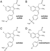
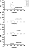
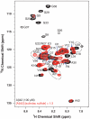
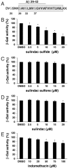
Following ectodomain shedding by beta-secretase, successive proteolytic cleavages within the transmembrane sequence (TMS) of the amyloid precursor protein (APP) catalyzed by gamma-secretase result in the release of amyloid-beta (Abeta) peptides of variable length. Abeta peptides with 42 amino acids appear to be the key pathogenic species in Alzheimer's disease, as they are believed to initiate neuronal degeneration. Sulindac sulfide, which is known as a potent gamma-secretase modulator (GSM), selectively reduces Abeta42 production in favor of shorter Abeta species, such as Abeta38. By studying APP-TMS dimerization we previously showed that an attenuated interaction similarly decreased Abeta42 levels and concomitantly increased Abeta38 levels. However, the precise molecular mechanism by which GSMs modulate Abeta production is still unclear. In this study, using a reporter gene-based dimerization assay, we found that APP-TMS dimers are destabilized by sulindac sulfide and related Abeta42-lowering compounds in a concentration-dependent manner. By surface plasmon resonance analysis and NMR spectroscopy, we show that sulindac sulfide and novel sulindac-derived compounds directly bind to the Abeta sequence. Strikingly, the attenuated APP-TMS interaction by GSMs correlated strongly with Abeta42-lowering activity and binding strength to the Abeta sequence. Molecular docking analyses suggest that certain GSMs bind to the GxxxG dimerization motif in the APP-TMS. We conclude that these GSMs decrease Abeta42 levels by modulating APP-TMS interactions. This effect specifically emphasizes the importance of the dimeric APP-TMS as a promising drug target in Alzheimer's disease.
Conflict of interest statement
The authors declare no conflict of interest.
Figures


Similar articles
-
Independent generation of Abeta42 and Abeta38 peptide species by gamma-secretase.J Biol Chem. 2008 Jun 20;283(25):17049-54. doi: 10.1074/jbc.M802912200. Epub 2008 Apr 21. J Biol Chem. 2008. PMID: 18426795
-
CTF1-51, a truncated carboxyl-terminal fragment of amyloid precursor protein, suppresses the effects of Aβ42-lowering γ-secretase modulators.Neurosci Lett. 2012 Sep 27;526(2):96-9. doi: 10.1016/j.neulet.2012.08.029. Epub 2012 Aug 23. Neurosci Lett. 2012. PMID: 22940081
-
Potent γ-secretase inhibitors/modulators interact with amyloid-β fibrils but do not inhibit fibrillation: a high-resolution NMR study.Biochem Biophys Res Commun. 2014 May 16;447(4):590-5. doi: 10.1016/j.bbrc.2014.04.041. Epub 2014 Apr 18. Biochem Biophys Res Commun. 2014. PMID: 24747079
-
γ-Secretase modulator in Alzheimer's disease: shifting the end.J Alzheimers Dis. 2012;31(4):685-96. doi: 10.3233/JAD-2012-120751. J Alzheimers Dis. 2012. PMID: 22710916 Review.
-
Alzheimer's disease.Subcell Biochem. 2012;65:329-52. doi: 10.1007/978-94-007-5416-4_14. Subcell Biochem. 2012. PMID: 23225010 Review.
Cited by
-
Second generation γ-secretase modulators exhibit different modulation of Notch β and Aβ production.J Biol Chem. 2012 Sep 21;287(39):32640-50. doi: 10.1074/jbc.M112.376541. Epub 2012 Jul 31. J Biol Chem. 2012. PMID: 22851182 Free PMC article.
-
Targeting the lateral interactions of transmembrane domain 5 of Epstein-Barr virus latent membrane protein 1.Biochim Biophys Acta. 2012 Sep;1818(9):2282-9. doi: 10.1016/j.bbamem.2012.05.013. Epub 2012 May 17. Biochim Biophys Acta. 2012. PMID: 22609737 Free PMC article.
-
Enzyme-linked immunosorbent assay-based method to quantify the association of small molecules with aggregated amyloid peptides.Anal Chem. 2012 Feb 7;84(3):1786-91. doi: 10.1021/ac2030859. Epub 2012 Jan 25. Anal Chem. 2012. PMID: 22243436 Free PMC article.
-
Development and mechanism of γ-secretase modulators for Alzheimer's disease.Biochemistry. 2013 May 14;52(19):3197-216. doi: 10.1021/bi400377p. Epub 2013 May 2. Biochemistry. 2013. PMID: 23614767 Free PMC article. Review.
-
Modulators of γ-secretase activity can facilitate the toxic side-effects and pathogenesis of Alzheimer's disease.PLoS One. 2013;8(1):e50759. doi: 10.1371/journal.pone.0050759. Epub 2013 Jan 7. PLoS One. 2013. PMID: 23308095 Free PMC article.
References
Publication types
MeSH terms
Substances
LinkOut - more resources
Full Text Sources
Other Literature Sources
Molecular Biology Databases

