. 2010 Oct 15;285(42):32638-46.
doi: 10.1074/jbc.M110.105544. Epub 2010 Aug 2.

WSS25 inhibits growth of xenografted hepatocellular cancer cells in nude mice by disrupting angiogenesis via blocking bone morphogenetic protein (BMP)/Smad/Id1 signaling

Affiliations

WSS25 inhibits growth of xenografted hepatocellular cancer cells in nude mice by disrupting angiogenesis via blocking bone morphogenetic protein (BMP)/Smad/Id1 signaling

Hong Qiu et al. J Biol Chem. .

Abstract

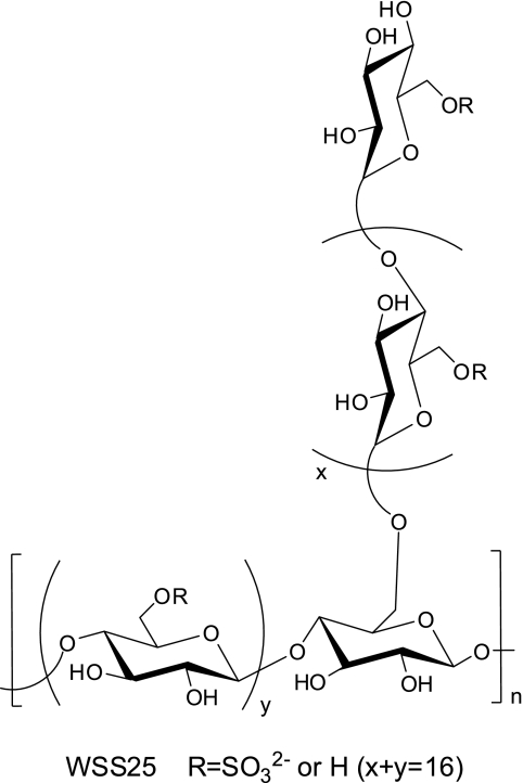
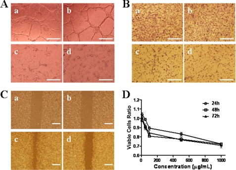
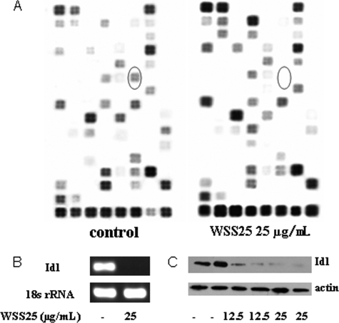
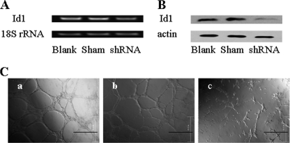
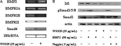
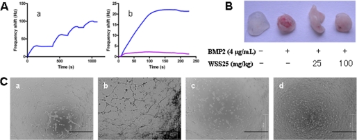
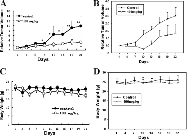
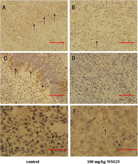
The highly expressed Id1 (inhibitor of DNA binding/differentiation) protein promotes angiogenesis in HCC and is a well established target for anti-angiogenesis therapeutic strategies. Heparan sulfate (HS) mimetics such as PI-88 can abrogate HS-protein interactions to inhibit angiogenesis. Id1 is the direct downstream effector of bone morphogenetic proteins (BMPs), which are angiogenic and HS-binding proteins. Thus, targeting BMPs by HS mimetics may inhibit angiogenesis via attenuating Id1 expression. We report here that a HS mimetic WSS25 potently inhibited the tube formation of HMEC-1 cells on Matrigel and their migration. Meanwhile, WSS25 (25 μg/ml) nearly completely blocked Id1 expression in the HMEC-1 cells as demonstrated by oligo-angiogenesis microarray analysis and further confirmed by RT-PCR and Western blotting. BMP/Smad/Id1 signaling also was blocked by WSS25 treatment in HMEC-1 cells. Importantly, Id1 knockdown in HMEC-1 cells caused the disruption of their tube formation on Matrigel. By employing quartz crystal microbalance analysis, we found that WSS25 strongly bound to BMP2. Moreover, WSS25 impaired BMP2-induced tube formation of HMEC-1 cells on Matrigel and angiogenesis in Matrigel transplanted into C57BL6 mice. Furthermore, WSS25 (100 mg/kg) abrogated the growth of HCC cells xenografted in male nude mice. Immunohistochemical analysis showed that both the expression of Id1 and the endothelial cell marker CD31 were lower in the WSS25-treated tumor tissue than in the control. Therefore, WSS25 is a potential drug candidate for HCC therapy as a tumor angiogenesis inhibitor.


Figures

FIGURE 1.
FIGURE 1.
Structural diagram of WSS25.
FIGURE 2.
FIGURE 2.
WSS25 impaired the tube formation of HMEC-1 cells on Matrigel and migration. A, WSS25 inhibited the tube formation of HMEC-1 cells on Matrigel. HMEC-1 cells (90 μl) treated with WSS25 (10 μl) at different final concentrations (panel b, 6.25 μg/ml; panel c, 25 μg/ml; panel d, 50 μg/ml); or vehicle (panel a) were seeded into the 96-well plate precoated with 50 μl Matrigel for 10 h. B, WSS25 impaired the migration of HMEC-1 cells in a trans-well migration assay. HMEC-1 cells were seeded into the inner chamber with MCDB131 medium containing 10 μg/ml (panel b), 100 μg/ml (panel c), 1 mg/ml WSS25 (panel d) or vehicle (panel a). C, WSS25 inhibited the migration of HMEC-1 cells in a wound healing assay (panels a and c, control; panels b and d, 25 μg/ml). D, HMEC-1 cells were seeded into the 96-well plate. After 24 h of incubation, WSS25 was added to the final concentrations of 1 μg/ml, 10 μg/ml, 100 μg/ml, 500 μg/ml, or 1 mg/ml. The cell viabilities were determined by the MTT assay 24 h (○), 48 h (□), or 72 h ([tric]) later. The results are representative of triplicate experiments.
FIGURE 3.
FIGURE 3.
WSS25 potently inhibited the expression of Id1. A, HMEC-1 cells were treated with WSS25 (25 μg/ml) for 18 h. RNA was then extracted for the oligo-angiogenesis microarray analysis. Circles indicate Id1. B, WSS25 at 25 μg/ml nearly completely inhibited the Id1 mRNA expression in HMEC-1 cells with 18 S rRNA as the internal control. C, WSS25 down-regulated the Id1 protein expression in HMEC-1 cells in a dose-dependent manner. β-Actin was used as a control for protein loading. Except for the microarray analysis, all experiments were repeated three times.
FIGURE 4.
FIGURE 4.
Id1 knockdown in HMEC-1 cells caused disruption of tube formation on Matrigel. A and B, HMEC-1 cells were plated into six-well plates for 24 h before transfection with Id1 shRNA for 30 h. Both RT-PCR and Western blotting were then used to detect Id1 expression. Id1 shRNA could down-regulate Id1 expression at mRNA level (A) and protein level (B). C, the tube formation of HMEC-1 cells on Matrigel was disrupted (panel c) after Id1 shRNA transfection, compared with the blank (panel a) and sham control (panel b). HMEC-1 cells (2.5 × 10⁵ cells/well) were seeded into a six-well plate for 24 h before transfection. The shRNA plasmids were transfected into the cells three times 24 h apart using X-fect polymer (Clontech). The cells were used in the tube formation assay as described under “Experimental Procedures” 24 h after the last transfection. Photos were taken after another 12 h of incubation. The results are representative of three experiments.
FIGURE 5.
FIGURE 5.
Effects of WSS25 on Id1, BMP2, and Smad signaling pathway components. A, WSS25 displayed different effects on the expressions of BMP2, BMPRIA, BMPRIB, BMPRII, and Smad4. RT-PCR analysis was performed as described under “Experimental Procedures.” B, WSS25 or Noggin inhibited both Smad1/5/8 phosphorylation and Id1 expression in HMEC-1 cells induced by BMP2. The cells were pretreated with 25 μg/ml of WSS25, 5 μg/ml noggin or vehicle for 23 h. The cells were then treated with 50 ng/ml of BMP2 or vehicle for another hour. The extracted proteins were analyzed by Western blotting using Id1, Smad1, and pSmad1/5/8 antibodies. β-Actin was used as a control for protein loading. The experiments were repeated twice.
FIGURE 6.
FIGURE 6.
Interaction of WSS25 with BMP2 and its effects on BMP2-induced angiogenesis. A, WSS25 strongly bound to BMP2 by QCM analysis. The WSS25 biosensor surface was prepared for measuring carbohydrate-protein interactions, where the frequency shift produced from biotinylated WSS25 binding to the streptavidin surface is shown in (panel a). The WSS25-BMP2 interaction was tested by injecting BMP2 (50 μg/ml, 50 μl) in running buffer onto the WSS25 biosensor surface, and the frequency response is displayed in panel b (blue curve). The large shift produced indicated that WSS25 bound to BMP2 strongly. As a control, the frequency responses by the BMP2 interaction with the streptavidin surface were measured, yielding a small response in panel b (pink curve). These results indicate the BMP2-WSS25 interaction was specific. B, WSS25 inhibited BMP2-induced angiogenesis in the Matrigel plug assay. Matrigel (500 μl) with or without BMP2 (4 μg/ml) was subcutaneously injected into the ventral region of C57/BL6 mice. WSS25 (25 mg/kg or 100 mg/kg body weight) was administered to the mice every other day from the second day after the Matrigel was plugged into the mice. Normal saline was used as the control. C, WSS25 and Noggin inhibited BMP2-induced tube formation of HMEC-1 cells on Matrigel. Growth factor reduced Matrigel (50 μl/well) was added to a 96-well plate to be solidified in 37 °C for 30 min. HMEC-1 cells (3 × 10⁴ cells in 98 μl MCDB131 medium supplemented with 0.1% FBS per well) were seeded into the 96-well plate after the Matrigel solidification. BMP2 (200 ng/ml) was added together with the cells only (panel b), or in the presence of Noggin (1 μg/ml) (panel c) or WSS25 (25 μg/ml) (panel d). Photos were taken after incubation for 24 h at 37 °C.
FIGURE 7.
FIGURE 7.
WSS25 represses the growth of HCC xenografted in nude mice. Bel7402 (A) and SMMC7721 (B) cells were subcutaneously injected into the front pad of male nude mice. For the Bel7402 xengografts (five mice/group), after the tumor volume grew to ∼100 mm3, 100 mg/kg of WSS25 in normal saline or the vehicle were injected via tail vein every other day. For the SMMC7721 xenografts, well developed tumors were cut into 1–3 mm3 fragments and transplanted subcutaneously into the right flank of the nude mice using a trocar under sterile conditions. When the tumor volume reached ∼100 mm3, the mice were randomly assigned into control and treatment groups (six mice/group), and the vehicle or 100 mg/kg WSS25 was administered via tail vein every other day. Tumor volume was measured using a caliber on the indicated days. Mice were sacrificed 22 days after treatment. The therapeutic effect of the compounds was expressed as the volume ratio of treatment to control (T/C). T/C (%) = 100% × (mean RTV of the treated group/mean RTV of the control group). The tumor growth was significantly inhibited in both tumor models. *, p < 0.05; **, p < 0.01. The T/C (%) for Bel7402 and SMMC7721 was 32.2 and 56%, respectively. The body weights of the mice showed no significant changes during the experiments.
FIGURE 8.
FIGURE 8.
Id1, phosphorylated Smad1/5/8, and CD31 expression levels were lower in WSS25-treated tumor tissue than in the control. Immunohistochemical analysis was performed on tumors from mice treated with 100 mg/kg of WSS25 via tail vein injection or the vehicle as described above. Compared with the control (A), there was also a reduction in the number of blood vessels (shown by reduced expression of the CD31 endothelial marker) in tumors treated with 100 mg/kg WSS25 (B) as indicated by the arrow. Representative images showed that treatment with WSS25 down-regulated Id1 expression in the tumor tissue (C) compared with the control (D) as indicated by the arrow. Phosphorylated Smad1/5/8 expression was also reduced in WSS25-treated tissues (F) compared with the control (E). All images are at 400× magnification.

References

    1. Tang Z. Y. (2001) World J. Gastroenterol. 7, 445–454 - PMC - PubMed
    1. Sun H. C., Tang Z. Y. (2004) J. Cancer Res. Clin. Oncol. 130, 307–319 - PMC - PubMed
    1. Perk J., Iavarone A., Benezra R. (2005) Nat. Rev. Cancer. 5, 603–614 - PubMed
    1. Ouyang X. S., Wang X., Lee D. T., Tsao S. W., Wong Y. C. (2002) J. Urol. 167, 2598–2602 - PubMed
    1. Perk J., Gil-Bazo I., Chin Y., de Candia P., Chen J. J., Zhao Y., Chao S., Cheong W., Ke Y., Al-Ahmadie H., Gerald W. L., Brogi E., Benezra R. (2006) Cancer Res. 66, 10870–10877 - PubMed

Publication types

MeSH terms