. 2010 Oct;78(10):4402-20.
doi: 10.1128/IAI.00398-10. Epub 2010 Aug 2.

Mucosal immunization with Vibrio cholerae outer membrane vesicles provides maternal protection mediated by antilipopolysaccharide antibodies that inhibit bacterial motility

Affiliations

Mucosal immunization with Vibrio cholerae outer membrane vesicles provides maternal protection mediated by antilipopolysaccharide antibodies that inhibit bacterial motility

Anne L Bishop et al. Infect Immun. 2010 Oct.

Abstract

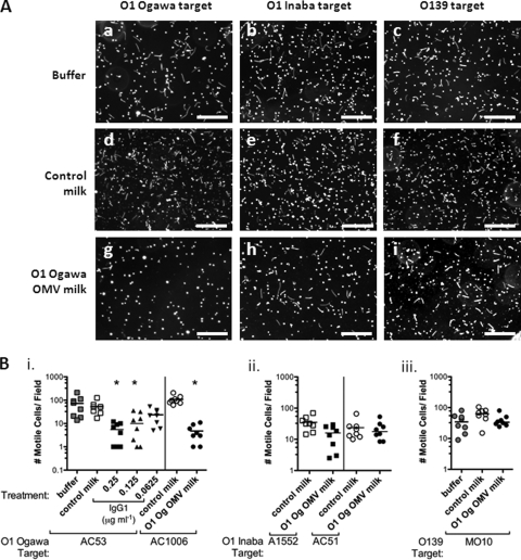
Vibrio cholerae is the causative agent of cholera, a severe diarrheal disease that remains endemic in many parts of the world and can cause outbreaks wherever sanitation and clean water systems break down. Prevention of disease could be achieved through improved sanitation and clean water provision supported by vaccination. V. cholerae serogroup O1 is the major cause of cholera; O1 serotypes Inaba and Ogawa have similar disease burdens, while O139 is the only non-O1 serogroup to cause epidemics. We showed previously that immunization of adult female mice with purified V. cholerae outer membrane vesicles (OMVs) elicits an antibody response that protect neonates from oral V. cholerae challenge and that suckling from an immunized dam accounts for the majority of protection from V. cholerae colonization. Here we report that lipopolysaccharide (LPS) is the major OMV protective antigen. Mucosal immunization with OMVs from Inaba or Ogawa provides significant cross-serotype protection from V. cholerae colonization, although serotype-specific antigens are dominant. OMVs from O1 or O139 do not provide cross-serogroup protection, but by immunization with a mixture of O1 and O139 OMVs, cross-serogroup protection was achieved. Neonatal protection is not associated with significant bacterial death but may involve inhibition of motility, as antibodies from OMV-immunized mice inhibit V. cholerae motility in vitro, with trends that parallel in vivo protection. Motility assays also reveal that a higher antibody titer is required to immobilize O139 compared to O1, a phenotype that is O139 capsule dependent.


Figures

FIG. 1.
FIG. 1.
V. cholerae OMV two-dose immunization and challenge. Immunization and challenge timeline using three doses (A) or two doses (B). (C) Anti-O1 Ogawa OMV ELISA probed with preimmune (day −1) and terminal serum (day 57) from mice immunized with O1 Ogawa OMVs using the two-dose protocol and anti-mouse IgG1 secondary antibody. Immunization route was either i.n. or oral as indicated. Each symbol represents serum from one mouse. *, P < 0.05 significantly above preimmune serum (Mann-Whitney U tests). (D) Small intestinal viable counts 24 h after challenge with ca. 105 O1 Ogawa V. cholerae CFU (exact input does were 0.7 to 0.9 ×10⁵) for neonates born to unimmunized (control) mice or mice immunized i.n. (i.n. OMVs) or orally (oral OMVs) with O1 Ogawa OMVs. Each symbol represents one neonate. *, P < 0.05, significant protection compared to controls is provided by i.n., but not oral, OMV immunization using a two-dose immunization protocol (Kruskal-Wallis and post hoc Dunn's multiple comparison tests). Bars, medians; dotted line, limit of detection.
FIG. 2.
FIG. 2.
OMV immunization elicits responses to conserved proteins, but proteinase K-resistant antigens are dominant. A total 5 μg of OMVs derived from V. cholerae O1 Ogawa strain AC53 (Og), O1 Inaba strain A1552 (In), O139 strain MO10 (O139) or otnA acapsular O139 mutant (otnA) were separated by SDS-PAGE (NuPAGE Bis-Tris 4 to 12% gels) and either silver stained (A) or transferred to nitrocellulose and probed with immune serum (day 99 bleeds) from mice i.n. immunized with O1 Ogawa OMVs (B). (C and D) Twenty-five-milligram samples of O1 Ogawa OMVs were kept frozen (lane 1) or incubated overnight (o/n) at 55°C alone (lane 2) or in the presence of 0.2% SDS (lane 3), 200 μg ml−1 proteinase K (lane 4), or 0.2% SDS and 200 μg ml−1 proteinase K (lane 5). (C) The OMVs were separated by SDS-PAGE and silver stained. (D) After each treatment, OMVs were diluted to 5 μg ml−1 and used to coat ELISA plates that were probed for binding of anti-O1 Ogawa OMV (day 38) serum samples and anti-IgG1 secondary antibody. Each symbol represents serum from one O1 Ogawa OMV-immunized mouse. Bars = medians. In panels A to C, arrows show the positions of the lipid A-plus-core polysaccharide LPS precursor (A-core) and full-length O1 or O139 LPS molecules after the addition of their terminal O antigens (A and C) and two conserved OMV protein antigens (Conserved) and anti-O1 LPS signal detected by Western blotting (B).
FIG. 3.
FIG. 3.
Antibody responses after immunization with O1 Ogawa, O1 Inaba, or O139 OMVs. Terminal bleed serum samples from mice immunized with O1 Ogawa (A), O1 Inaba (B), a mixture of O1 Ogawa + Inaba (C), or O139 (D) OMVs by the i.n. or oral route, as indicated, were tested for binding to O1 Ogawa (Og), O1 Inaba (In), or O139 OMV-coated ELISA plates, as indicated, and anti-IgG1 secondary antibody. In panels A to C, *, P < 0.05, significant differences in binding between O1 Ogawa and Inaba OMVs (Kruskal-Wallis and posthoc Dunn's multiple comparison tests). n/s, not significant. (D) *, P < 0.05, binding of anti-O139 OMV serum was significantly greater for O139 OMVs than either O1 Inaba or O1 Ogawa OMVs (Kruskal-Wallis and post hoc Dunn's multiple comparison tests). Each symbol represents serum from one mouse. Bars, medians; dotted line, limit of detection.
FIG. 4.
FIG. 4.
Homologous and heterologous V. cholerae challenge of neonates born to mice immunized with O1 or O139 OMVs. Mice were immunized by i.n. or oral route with either O1 Ogawa (Og), O1 Inaba (In), a mixture of O1 Ogawa + Inaba (Og + In), or O139 OMVs, as indicated. Control mice were sham-immunized with PBS by i.n. or oral route. An immunization protocol with three doses and 25 μg of OMVs per dose was used. Mice were mated, and their neonates were challenged at ca. 500× the ID50 with O1 Ogawa AC53 (A), O1 Inaba A1552 (B), or O139 MO10 (C). Exact input doses were as follows: O1 Ogawa, 6.7 ×10⁴ to 2.1 ×10⁵ CFU; O1 Inaba, 2.6 to 9.7 ×10⁴ CFU; and O139, 3.5 to 8.1 ×10⁵ CFU. Each symbol represents small-intestinal viable counts for one neonate 24 h postinfection. *, P < 0.05, compared to controls sham immunized via the same route (Kruskal-Wallis and post hoc Dunn's multiple comparison tests). Bars, medians; dotted line, limit of detection.
FIG. 5.
FIG. 5.
Spatial and temporal distribution of V. cholerae viable counts in neonates from OMV-immunized and control mice. (A) Viable counts for V. cholerae in the small intestine (SI) or large intestine (LI) 2 or 4 h postinfection as indicated for neonates born to PBS control-immunized (closed symbols) or O1 Ogawa OMV-immunized (open symbols) dams. (B) Viable V. cholerae counts in the stomach (ST), SI, or LI or total counts 1 h postinfection for neonates born to sham-immunized (Control, closed symbols) or O1 Ogawa OMV-immunized (OMV, open symbols) immunized dams. *, P < 0.05; +, P = 0.042, Mann-Whitney U tests, control compared with OMV-immunized mice. Each symbol represents one neonate. Bars, medians; dotted line, limit of detection.
FIG. 6.
FIG. 6.
Milk from O1 Ogawa OMV-immunized mice specifically inhibits O1 Ogawa motility. Buffer alone or milk from neonates born to i.n. sham-immunized (control milk) or O1 Ogawa OMV-immunized mice (O1 Og OMV milk) was mixed 1:2 with V. cholerae (OD600 = 0.05) in LB. After 10 min, bacterial motility was observed by dark-field microscopy using a 400-ms exposure. The number of motile bacteria per field, which appear as lines or swirls, were counted. (A) Examples of images showing O1 Ogawa AC53 (a, d, and g), O1 Inaba A1552 (b, e, and h), or O139 MO10 (c, f, and i) targets mixed with buffer alone (a, b, and c), control milk (d, e, and f), or O1 Ogawa OMV-immunized mouse milk (g, h, and i). Scale bars equal 100 μm. (B) Quantification of motility in the presence of buffer, control milk, or O1 Ogawa OMV-immune milk, as indicated, for O1 Ogawa strains AC53 or AC1006 (i); O1 Inaba strains A1552 or AC51 (ii); O139 strain MO10 (iii). (Bi) For the O1 Ogawa AC53 target, different dilutions of immune milk were made to give 0.25, 0.125, or 0.0625 μg ml−1 of anti-O1 Ogawa (Og) OMV IgG1. In all other cases, immune milk was diluted to give 0.25 μg ml−1 IgG1, and control milk was diluted 1 in 1.6 (the lowest dilution used for immune milk). Each symbol represents milk from one mouse, or one buffer control, mixed with the indicated target bacterial suspension. *, P < 0.05, significant inhibition of motility compared to control milk (Kruskal-Wallis and Dunn's multiple comparison tests). Bars = medians.
FIG. 7.
FIG. 7.
O139 capsule increases the amount of antibody required to inhibit motility. Serum from control sham-immunized (diluted 1 in 1.1, the lowest dilution needed for the immune serum), O139 OMV-immunized, or O139 + O1 OMV-immunized mice at 0.25, 1, or 4 μg ml−1 of anti-OMV IgG1, as indicated, was mixed 1:2 with either wild-type O139 MO10 (A), acapsular O139 mutant MO10 otnA::pGP704 (B), or O1 Ogawa AC53 (C) targets each diluted in LB to an OD600 of 0.05. After 10 min, motility was observed by dark-field microscopy with a 400-ms exposure. Numbers of motile cells per field were counted. Each symbol represents motility of the indicated target bacterium in the presence of serum from one mouse. *, P < 0.05, significant inhibition of motility compared to control serum (Kruskal-Wallis and Dunn's multiple comparison tests). Bars, medians; dotted line, limit of detection.
FIG. 8.
FIG. 8.
Immunization with a mixture of O1 and O139 OMVs provides cross-serogroup protection. Mice were immunized i.n. with a 1:1:1 mixture of O1 Ogawa + O1 Inaba + O139 OMVs using 25 μg total OMVs per dose and a three-dose immunization. (A) Terminal bleeds from these mice show IgG1 (i), IgA (ii), and total Ig (iii) responses, measured by ELISA and vibriocidal antibody titers (iv) against O1 Ogawa (Og), O1 Inaba (In), or O139 OMVs (ELISAs) or live bacteria (vibriocidal assay), as indicated, that were similar for O1 Ogawa, Inaba, and O139. Each symbol represents serum from one immunized mouse. (B) Neonates born to these mice were protected from small intestinal colonization (viable counts), compared to control sham-immunized mice, upon challenge with O1 Ogawa AC53 (input 1.2 to 2.2 ×10⁵) (i), O1 Inaba A1552 (input 5.9 to 6.9 ×10⁴) (ii), or O139 (input 2.8 to 7.6 ×10⁵) (iii). Each symbol represents one infected neonate born to control or O1 Ogawa + O1 Inaba + O139 OMV-immunized mice as indicated. *, P < 0.05, significantly lower viable counts compared to control (Mann-Whitney U test). Bars, medians; dotted line, limit of detection.

Similar articles

Cited by

References

    1. Ahmed, T., A. M. Svennerholm, A. Al Tarique, G. N. Sultana, and F. Qadri. 2009. Enhanced immunogenicity of an oral inactivated cholera vaccine in infants in Bangladesh obtained by zinc supplementation and by temporary withholding breast-feeding. Vaccine 27:1433-1439. - PubMed
    1. Alaniz, R. C., B. L. Deatherage, J. C. Lara, and B. T. Cookson. 2007. Membrane vesicles are immunogenic facsimiles of Salmonella typhimurium that potently activate dendritic cells, prime B and T cell responses, and stimulate protective immunity in vivo. J. Immunol. 179:7692-7701. - PubMed
    1. Albert, M. J., K. Alam, M. Ansaruzzaman, F. Qadri, and R. B. Sack. 1994. Lack of cross-protection against diarrhea due to Vibrio cholerae O139 (Bengal strain) after oral immunization of rabbits with V. cholerae O1 vaccine strain CVD103-HgR. J. Infect. Dis. 169:230-231. - PubMed
    1. Albert, M. J., K. Alam, A. S. Rahman, S. Huda, and R. B. Sack. 1994. Lack of cross-protection against diarrhea due to Vibrio cholerae O1 after oral immunization of rabbits with V. cholerae O139 Bengal. J. Infect. Dis. 169:709-710. - PubMed
    1. Albert, M. J., A. K. Siddique, M. S. Islam, A. S. Faruque, M. Ansaruzzaman, S. M. Faruque, and R. B. Sack. 1993. Large outbreak of clinical cholera due to Vibrio cholerae non-O1 in Bangladesh. Lancet 341:704. - PubMed

Publication types