. 2010 Oct;78(10):4134-50.
doi: 10.1128/IAI.00167-10. Epub 2010 Aug 2.

Three Yersinia pestis adhesins facilitate Yop delivery to eukaryotic cells and contribute to plague virulence

Affiliations

Three Yersinia pestis adhesins facilitate Yop delivery to eukaryotic cells and contribute to plague virulence

Suleyman Felek et al. Infect Immun. 2010 Oct.

Abstract

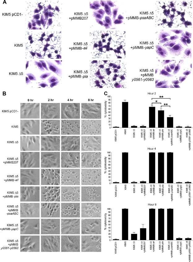
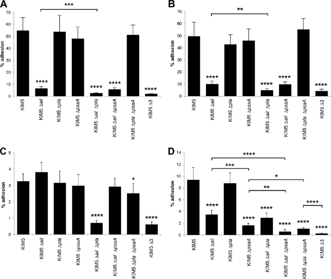
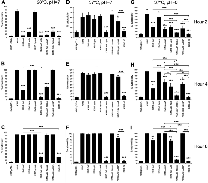
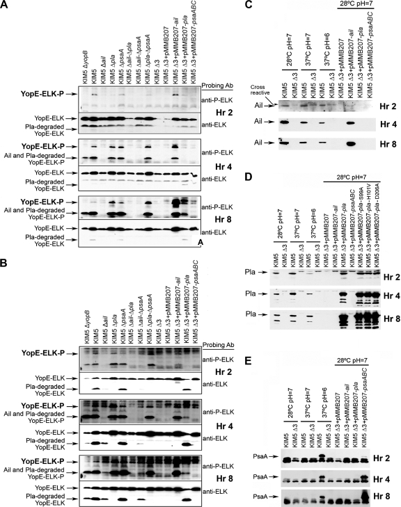
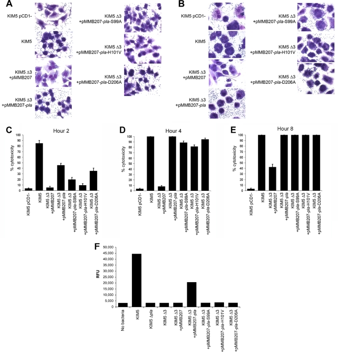
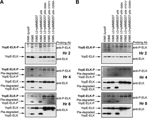
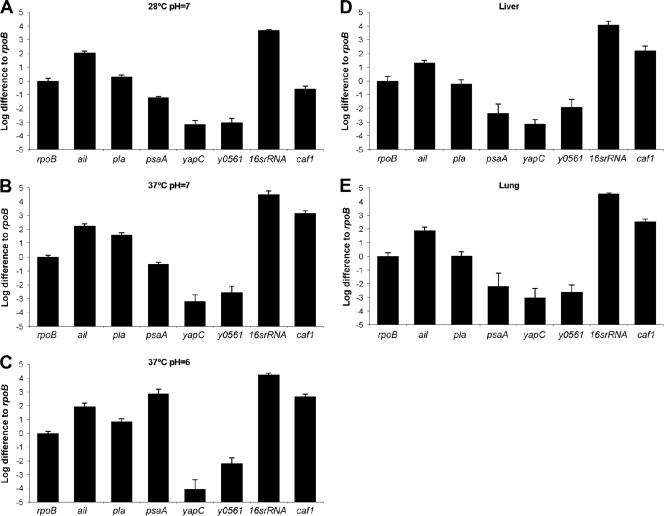
To establish a successful infection, Yersinia pestis requires the delivery of cytotoxic Yops to host cells. Yops inhibit phagocytosis, block cytokine responses, and induce apoptosis of macrophages. The Y. pestis adhesin Ail facilitates Yop translocation and is required for full virulence in mice. To determine the contributions of other adhesins to Yop delivery, we deleted five known adhesins of Y. pestis. In addition to Ail, plasminogen activator (Pla) and pH 6 antigen (Psa) could mediate Yop translocation to host cells. The contribution of each adhesin to binding and Yop delivery was dependent upon the growth conditions. When cells were pregrown at 28°C and pH 7, the order of importance for adhesins in cell binding and cytotoxicity was Ail > Pla > Psa. Y. pestis grown at 37°C and pH 7 had equal contributions from Ail and Pla but an undetectable role for Psa. At 37°C and pH 6, both Ail and Psa contributed to binding and Yop delivery, while Pla contributed minimally. Pla-mediated Yop translocation was independent of protease activity. Of the three single mutants, the Δail mutant was the most defective in mouse virulence. The expression level of ail was also the highest of the three adhesins in infected mouse tissues. Compared to an ail mutant, additional deletion of psaA (encoding Psa) led to a 130,000-fold increase in the 50% lethal dose for mice relative to that of the KIM5 parental strain. Our results indicate that in addition to Ail, Pla and Psa can serve as environmentally specific adhesins to facilitate Yop secretion, a critical virulence function of Y. pestis.


Figures

FIG. 1.
FIG. 1.
Adhesion and cytotoxicity of the KIM5 Δ5 mutant complemented with various Y. pestis adhesins. (A) Giemsa-stained infection assays at 2 h postinoculation. Cell rounding and bound bacteria are visible. Pictures are representative of overall effects. (B) Cell rounding/cytotoxicity assay of KIM5 derivatives, visualized by phase-contrast microscopy. HEp-2 cells were infected, and cell rounding was observed at hours 2, 4, and 8. (C) Quantification of cell rounding data from panel B at various time points (5 fields/strain; n, ∼500 cells). As a negative control, a KIM5 strain cured of the Yop-encoding virulence plasmid, pCD1, was included. *, P < 0.05; **, P < 0.000001. Error bars represent standard deviations.
FIG. 2.
FIG. 2.
Cell adhesion of various KIM5 mutant derivatives. Binding to HEp-2 cells (A) or THP-1 cells (B) by various KIM5 mutants grown at 28°C and pH 7 was assessed using a CFU assay. THP-1 cells were pretreated with 5 μg/ml of cytochalasin D to prevent phagocytosis during the cell adhesion assays. Binding to HEp-2 cells was also assessed after growing strains at 37°C and pH 7 (C) or at 37°C and pH 6 (D). Δ3 denotes KIM5 Δail Δpla ΔpsaA. *, P < 0.05; **, P < 0.005; ***, P < 0.0005; ****, P < 0.00005. Error bars represent standard deviations.
FIG. 3.
FIG. 3.
Cytotoxicity time course for HEp-2 cells. Y. pestis KIM5 and mutant derivatives were added to HEp-2 cells, and cell rounding was observed at hours 2 (A, D, and G), 4 (B, E, and H), and 8 (C, F, and I). Strains were grown at 28°C and pH 7 (A to C), 37°C and pH 7 (D to F), or 37°C and pH 6 (G to I) prior to infection of cells. Cytotoxicity was quantified by counting rounded cells in 5 fields/strain (n, ∼500 cells). Δ3 denotes KIM5 Δail Δpla ΔpsaA. *, P < 0.05; **, P < 0.00005; ***, P < 0.000005. Error bars represent standard deviations.
FIG. 4.
FIG. 4.
Yop delivery to eukaryotic cells. Yop translocation was measured by assessing delivery to and phosphorylation of ELK-tagged YopE from Y. pestis KIM5 and derivative strains in HEp-2 (A) and THP-1 (B) cells. Infected cells were harvested at 2, 4, and 8 postinoculation, and extracts were analyzed by SDS-PAGE and Western blotting, using anti-phospho-ELK and anti-ELK antibodies. Extracts were also tested by Western blotting for Ail (C), Pla (D), and Psa (E) expression from an IPTG-inducible plasmid under various growth conditions over 8 h. A KIM5 ΔyopB strain (lacking a component of the Yop translocation apparatus) was included as a negative control. Δ3 denotes KIM5 Δail Δpla ΔpsaA.
FIG. 5.
FIG. 5.
Adhesion and cytotoxicity of Pla and nonproteolytic Pla mutants to HEp-2 (A) and THP-1 (B) cells after 2 h of incubation. (C to E) Time course quantification of cell rounding for HEp-2 cells. (F) Fluorescence cleavage assay for detection of Pla proteolytic activity. RFU, relative fluorescence units. Δ3 denotes KIM5 Δail Δpla ΔpsaA. Error bars represent standard deviations.
FIG. 6.
FIG. 6.
Yop translocation to HEp-2 and THP-1 cells by Pla mutants defective for proteolysis. HEp-2 (A) and THP-1 (B) cells were infected with KIM5 derivatives, and Western immunoblotting was performed for total ELK and phosphorylated ELK at the indicated time points. Δ3 denotes KIM5 Δail Δpla ΔpsaA.
FIG. 7.
FIG. 7.
Transcription levels of ail, pla, psaA, yapC, y0561, 16S rRNA gene, and caf1. Expression levels were determined in vitro under different growth conditions (A to C) and in vivo in livers (D) and lungs (E). All values are presented relative to rpoB expression. For in vivo samples, mice were infected i.v. with 960 CFU Y. pestis KIM5 and then sacrificed on day 4, and qRT-PCR was performed on liver and lung specimens. The colonization levels for tissues used for qRT-PCR were as follows: for liver tissue, 13,000,000 CFU/g; and for lung tissue, 3,000,000 CFU/g. Ten milligrams of tissue was used to generate mRNA for liver and lung tissues. Error bars represent standard deviations.

References

    1. Anisimov, A. P., I. V. Bakhteeva, E. A. Panfertsev, T. E. Svetoch, T. B. Kravchenko, M. E. Platonov, G. M. Titareva, T. I. Kombarova, S. A. Ivanov, A. V. Rakin, K. K. Amoako, and S. V. Dentovskaya. 2009. The subcutaneous inoculation of pH 6 antigen mutants of Yersinia pestis does not affect virulence and immune response in mice. J. Med. Microbiol. 58:26-36. - PubMed
    1. Bartra, S. S., K. L. Styer, D. M. O'Bryant, M. L. Nilles, B. J. Hinnebusch, A. Aballay, and G. V. Plano. 2008. Resistance of Yersinia pestis to complement-dependent killing is mediated by the Ail outer membrane protein. Infect. Immun. 76:612-622. - PMC - PubMed
    1. Bearden, S., J. Fetherston, and R. Perry. 1997. Genetic organization of the yersiniabactin biosynthetic region and construction of avirulent mutants in Yersinia pestis. Infect. Immun. 65:1659-1668. - PMC - PubMed
    1. Ben-Efraim, S., M. Aronson, and L. Bichowsky-Slomnicki. 1961. New antigenic component of Pasteurella pestis formed under specified conditions of pH and temperature. J. Bacteriol. 81:704-714. - PMC - PubMed
    1. Bichowsky-Slomnicki, L., and S. Ben-Efraim. 1963. Biological activities in extracts of Pasteurella pestis and their relation to the “pH 6 antigen.” J. Bacteriol. 86:101-111. - PMC - PubMed

Publication types