. 2010 Oct;30(19):4564-74.
doi: 10.1128/MCB.00146-10. Epub 2010 Aug 2.

The type I interferon signaling pathway is a target for glucocorticoid inhibition

Affiliations

The type I interferon signaling pathway is a target for glucocorticoid inhibition

Jamie R Flammer et al. Mol Cell Biol. 2010 Oct.

Abstract

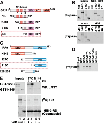
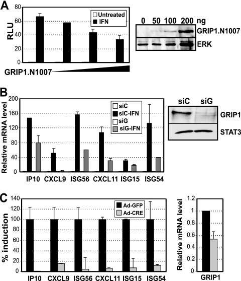
Type I interferon (IFN) is essential for host defenses against viruses; however, dysregulated IFN signaling is causally linked to autoimmunity, particularly systemic lupus erythematosus. Autoimmune disease treatments rely on glucocorticoids (GCs), which act via the GC receptor (GR) to repress proinflammatory cytokine gene transcription. Conversely, cytokine signaling through cognate Jak/STAT pathways is reportedly unaffected or even stimulated by GR. Unexpectedly, we found that GR dramatically inhibited IFN-stimulated gene (ISG) expression in macrophages. The target of inhibition, the heterotrimeric STAT1-STAT2-IRF9 (ISGF3) transcription complex, utilized the GR cofactor GRIP1/TIF2 as a coactivator. Consequently, GRIP1 knockdown, genetic ablation, or depletion by GC-activated GR attenuated ISGF3 promoter occupancy, preinitiation complex assembly, and ISG expression. Furthermore, this regulatory loop was restricted to cell types such as macrophages expressing the GRIP1 protein at extremely low levels, and pharmacological disruption of the GR-GRIP1 interaction or transient introduction of GRIP1 restored RNA polymerase recruitment to target ISGs and the subsequent IFN response. Thus, type I IFN is a cytokine uniquely controlled by GR at the levels of not only production but also signaling through antagonism with the ISGF3 effector function, revealing a novel facet of the immunosuppressive properties of GCs.


Figures

FIG. 1.
FIG. 1.
Dex inhibits IFN-induced gene expression downstream of Jak/STAT pathway activation. (A) Inhibition of IFN-induced gene expression by Dex. BMMΦ were treated for 1 h (ISG56, ISG15, ISG54, OASL1, Mx1), 2 h (IP10, CXCL9, CXCL11, IL-6), or 4 h (Rantes) with vehicle (untreated) or 500 U/ml IFN with or without 100 nM Dex, as shown. mRNA abundance of ISGF3 target genes was determined by qPCR, with GAPDH as the normalization control, and expressed relative to untreated cells (control = 1). Error bars represent ±standard errors of the mean (SEM). Results are from at least eight independent experiments. (B and C) Type I IFN-induced phosphorylation of STAT1 and STAT2 is Dex resistant. RAW 264.7 cells or BMMΦ were cultured for the indicated times in the presence of 500 U/ml IFN with (+) or without (−) 100 nM Dex. STAT1 and STAT2 expression and activation by tyrosine (Y701 and Y690, respectively) or serine (S727, STAT1 only) phosphorylation was assessed by immunoblotting.
FIG. 2.
FIG. 2.
IFN induction and Dex inhibition are mediated by ISREs. (A) Diagram of a series of IP10-derived luciferase reporters, with WT or mutated (stars) promoter elements and a dimerized, IFN-β-derived ISRE reporter (2xISRE). (B and C) CV-1 cells (105) were transfected with 200 ng pCDNA3.rGR, 35 ng pCMV-LacZ, and 200 ng of the indicated reporter constructs listed in panel A and treated the following day for 6 h as indicated. Luciferase activity was normalized to β-galactosidase activity (as a measure of transfection efficiency) and expressed as relative luminescence units (RLU). Error bars represent ±SEM. Results are from five independent experiments.
FIG. 3.
FIG. 3.
Dex inhibits IFN-induced transcription complex assembly. BMMΦ were treated as indicated for 30 min. ChIP assays were performed using antibodies to STAT1 (A), H3AcK9/K14 (B), Pol2 (C), or the isotype-matched control IgG (A to C). Occupancy was determined by qPCR amplification over ISRE (A) or TSS (B and C) regions of the indicated target genes, normalized to the internal control (45S), and expressed relative to the mean signal obtained from cells precipitated with control IgG (set to 1). Error bars represent ±SEM. Results are from at least three independent experiments.
FIG. 4.
FIG. 4.
GRIP1 and IRF9 interact in vitro in a GR-sensitive manner. (A and C) Domain diagrams of full-length GRIP1 (A) and IRF9 (C) and their derivatives produced in vitro and recombinantly in E. coli as GST fusion proteins, respectively. (B and D) Mapping the interacting surface on GRIP1 and IRF9. (B) 35S-radiolabeled GRIP1 derivatives listed in panel A were tested for their ability to interact with full-length recombinant GST-IRF9 (top) or GST alone (bottom). (D) Binding assays were performed between 35S-GRIP1 2-RD and GST-IRF9 derivatives listed in panel C. (E) The GRIP1-IRF9 interaction is disrupted by GR. His-tagged GRIP1 3-RD immobilized on affinity resin was incubated with GST-IRF9 127C (lanes 3 and 4) or N145 (lanes 5 and 6), in the presence or absence of 35S-GR, as indicated. Dex (1 μM) was present in all reactions. GR binding to 3-RD was verified by autoradiography (middle), and IRF9 binding was assessed by immunoblotting with GST-specific antibodies (top). Immobilized 3-RD was visualized by Coomassie blue staining (bottom).
FIG. 5.
FIG. 5.
IFN-induced gene expression is dependent upon the presence of active GRIP1. (A) GRIP1.N1007 overexpression attenuates IFN-induced transcription. CV-1 cells (105) were transiently transfected with 35 ng pCMV-LacZ, 200 ng 2xISRE-Luc, and increasing amounts (0, 50, 100, and 200 ng) of pCDNA GRIP1.N1007 or with pCDNA3 to equalize the total amount of transfected DNA. The following day, cells were treated for 6 h with 500 U/ml IFN, and whole-cell lysates were assayed for luciferase activity (exactly as described for Fig. 2) (left) or GRIP1 expression by immunoblotting (right). (B) siRNA depletion of GRIP1 antagonizes IFN-dependent ISG induction. RAW 264.7 cells (2 × 10⁶) were transfected with 3 μg of siRNA against GRIP1 (siG) or scrambled RNA (siC) as a negative control. Eighteen hours later, cells were treated with 500 U/ml IFN for 6 h. The GRIP1 protein level was analyzed by immunoblotting with anti-STAT3 blot to verify equal loading (right), and mRNA expression levels of target genes were analyzed by qPCR (left), as described for Fig. 1. (C) Adenovirus-mediated GRIP1 knockdown in primary MΦ attenuates the IFN response. Primary BMMΦ were derived from GRIP1flox/flox mice as described in Materials and Methods and infected with adenovirus-expressing Cre recombinase (Ad-Cre) or the control GFP (Ad-GFP). mRNA levels of the indicated genes were analyzed by qPCR, as described for Fig. 1. ISGs are expressed as a percentage of IFN induction in Ad-GFP-infected cells (100%). GRIP1 is expressed relative to the mean signal obtained from cells infected with the control Ad-GFP (set to 1). Error bars represent ±SEM. Results are from at least four independent experiments.
FIG. 6.
FIG. 6.
RU486 relieves Dex-mediated inhibition of ISG transcription. (A) BΜΜΦ were treated for 2 h with 500 U/ml IFN with or without 100 nM Dex, with or without the indicated concentrations of RU486 (RU). mRNA abundance of ISGF3 target genes was determined by qPCR, with β-actin as the normalization control, and is expressed as a percentage of induction by IFN alone (100%). (B) BMMΦ were treated as indicated for 30 min. ChIP assays were performed using Pol2 antibodies or isotype-matched control normal IgG (not shown). Occupancy was determined by qPCR amplification over TSS regions of the indicated target genes, as described for Fig. 3. Error bars represent ±SEM.
FIG. 7.
FIG. 7.
The effect of glucocorticoids on ISG expression depends on the GRIP1 protein level in a cell. (A) ISG expression in 3T3 mouse fibroblasts is Dex resistant. 3T3 cells were treated for the indicated times with 500 U/ml IFN with or without 100 nM Dex (left) or Dex alone (right), and the mRNA abundance of the indicated genes was determined by qPCR, with β-actin as the normalization control. (B) GRIP1 protein level varies dramatically between cell types. An equivalent amount of whole-cell extracts (WCE) from BMMΦ, RAW 264.7 cells, or 3T3 fibroblasts were fractionated by SDS-PAGE, and the expression of GRIP1, GR, and ERK1/2 (as a loading control) was assessed by immunoblotting. (C) IFN-dependent GRIP1 and Pol2 recruitment to ISGs in fibroblasts is Dex resistant. 3T3 cells were treated for 1 h as indicated, and GRIP1 and Pol2 occupancy at the ISRE or TSS regions, respectively, of indicated genes was determined by qPCR, normalized to the internal control (45S), and expressed relative to the mean signal obtained from cells precipitated with the control IgG (set to 1). (D) GRIP1-IRF9 interaction in RAW 264.7 cells is sensitive to Dex. RAW 264.7 cells were treated as shown for 1 h, and lysates were prepared. Twenty percent of each lysate was boiled in sample buffer to generate WCE, whereas the rest was precipitated with anti-GRIP1 antibody (αGRIP1 IP). Protein complexes were adsorbed on protein A/G Plus agarose beads, boiled in sample buffer, and separated by SDS-PAGE, along with WCE. GRIP1 and IRF9 were detected by immunoblotting. (E) GRIP1 overexpression in RAW 264.7 cells rescues ISG expression. RAW 264.7 cells (0.5 × 10⁶) were transfected with 1 to 2 μg of pCDNA-GRIP1 (GRIP1) or empty vector (vec) using GenePORTER 3000 (Genlantis) as per the manufacturer's instructions. Cells were treated with IFN with or without Dex 24 h later for 2 h, and mRNA expression levels of IP10 and OASL1 were analyzed by qPCR, as described for Fig. 1.

Similar articles

Cited by

References

    1. Abraham, S. M., T. Lawrence, A. Kleiman, P. Warden, M. Medghalchi, J. Tuckermann, J. Saklatvala, and A. R. Clark. 2006. Antiinflammatory effects of dexamethasone are partly dependent on induction of dual specificity phosphatase 1. J. Exp. Med. 203:1883-1889. - PMC - PubMed
    1. Adelman, K., M. A. Kennedy, S. Nechaev, D. A. Gilchrist, G. W. Muse, Y. Chinenov, and I. Rogatsky. 2009. Immediate mediators of the inflammatory response are poised for gene activation through RNA polymerase II stalling. Proc. Natl. Acad. Sci. U. S. A. 106:18207-18212. - PMC - PubMed
    1. Aittomaki, S., M. Pesu, B. Groner, O. A. Janne, J. J. Palvimo, and O. Silvennoinen. 2000. Cooperation among Stat1, glucocorticoid receptor, and PU.1 in transcriptional activation of the high-affinity Fc gamma receptor I in monocytes. J. Immunol. 164:5689-5697. - PubMed
    1. An, J., R. C. Ribeiro, P. Webb, J. A. Gustafsson, P. J. Kushner, J. D. Baxter, and D. C. Leitman. 1999. Estradiol repression of tumor necrosis factor-alpha transcription requires estrogen receptor activation function-2 and is enhanced by coactivators. Proc. Natl. Acad. Sci. U. S. A. 96:15161-15166. - PMC - PubMed
    1. Baechler, E. C., F. M. Batliwalla, G. Karypis, P. M. Gaffney, W. A. Ortmann, K. J. Espe, K. B. Shark, W. J. Grande, K. M. Hughes, V. Kapur, P. K. Gregersen, and T. W. Behrens. 2003. Interferon-inducible gene expression signature in peripheral blood cells of patients with severe lupus. Proc. Natl. Acad. Sci. U. S. A. 100:2610-2615. - PMC - PubMed

Publication types

MeSH terms