. 2010 Oct;30(19):4687-97.
doi: 10.1128/MCB.01581-09. Epub 2010 Aug 2.

VRK2 inhibits mitogen-activated protein kinase signaling and inversely correlates with ErbB2 in human breast cancer

Affiliations

VRK2 inhibits mitogen-activated protein kinase signaling and inversely correlates with ErbB2 in human breast cancer

Isabel F Fernández et al. Mol Cell Biol. 2010 Oct.

Abstract

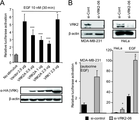
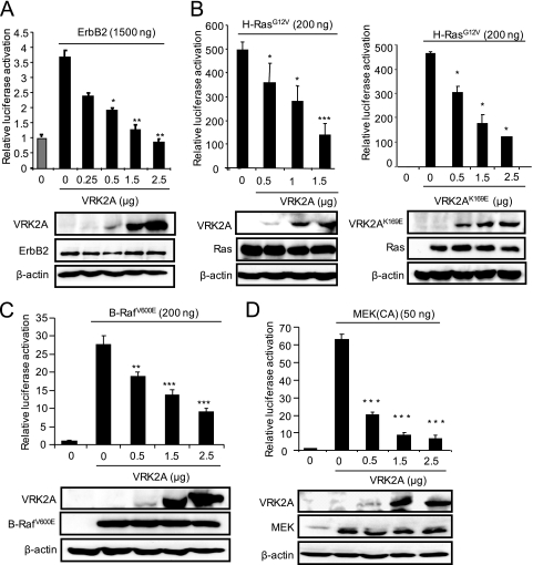
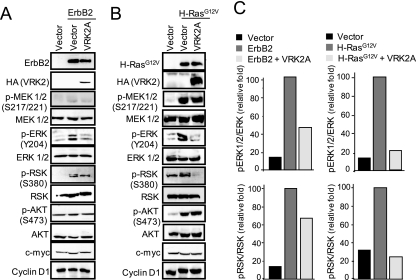
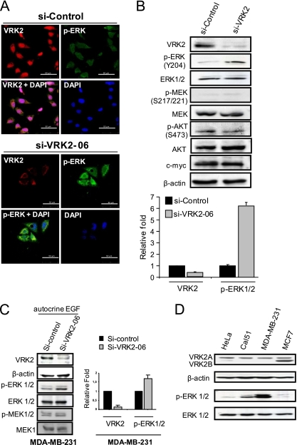
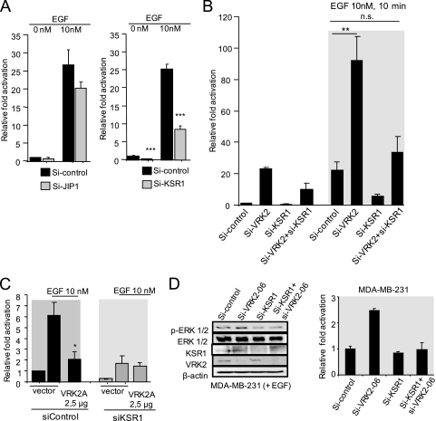
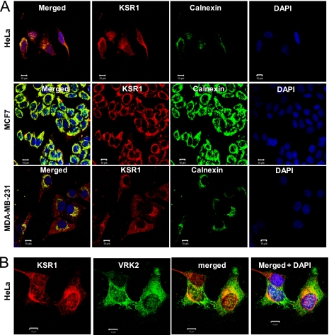
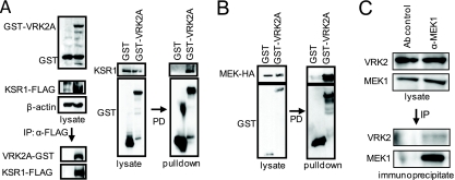
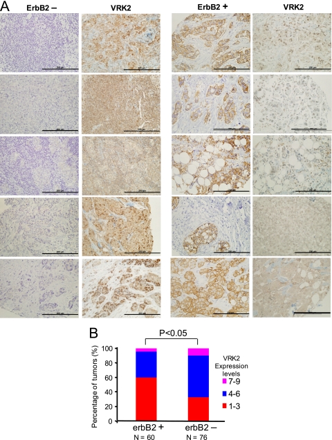
The epidermal growth factor (EGF)-ErbB-mitogen-activated protein kinase (MAPK) transcription signaling pathway is altered in many types of carcinomas, and this pathway can be regulated by new protein-protein interactions. Vaccinia-related kinase (VRK) proteins are Ser-Thr kinases that regulate several signal transduction pathways. In this work, we study the effect of VRK2 on MAPK signaling using breast cancer as a model. High levels of VRK2 inhibit EGF and ErbB2 activation of transcription by the serum response element (SRE). This effect is also detected in response to H-Ras(G12V) or B-Raf(V600E) oncogenes and is accompanied by a reduction in phosphorylated extracellular signal-regulated kinase (ERK) levels, p90RSK levels, and SRE-dependent transcription. Furthermore, VRK2 knockdown has the opposite effect, increasing the transcriptional response to stimulation with EGF and leading to increased levels of ERK phosphorylation. The molecular mechanism lies between MAPK/ERK kinase (MEK) and ERK, since MEK remains phosphorylated while ERK phosphorylation is blocked by VRK2A. This inhibition of the ERK signaling pathway is a consequence of a direct protein-protein interaction between VRK2A, MEK, and kinase suppressor of Ras 1 (KSR1). Identification of new correlations in human cancer can lead to a better understanding of the biology of individual tumors. ErbB2 and VRK2 protein levels were inversely correlated in 136 cases of human breast carcinoma. In ErbB2(+) tumors, there is a significant reduction in the VRK2 level, suggesting a role for VRK2A in ErbB2-MAPK signaling. Thus, VRK2 downregulation in carcinomas permits signal transmission through the MEK-ERK pathway without affecting AKT signaling, causing a signal imbalance among pathways that contributes to the phenotype of breast cancer.


Figures

FIG. 1.
FIG. 1.
VRK2A protein modulates EGF activation of SRE-dependent transcription. (A) Effect of VRK2A overexpression. MCF7 cells were transfected with 1 μg of the SRE-luciferase reporter, as well as plasmids, for overexpression of VRK2A or VRK1. The immunoblot shows the correct expression of the corresponding VRK proteins detected with an antibody to the HA epitope. The error bars indicate standard deviations. ***, P < 0.001; *, P < 0.05. (B) (Top) Knockdown of the VRK2 intracellular protein level with siVRK2-06 in MDA-MB-231 breast cancer cells and HeLa cells. MDA-MB-231 cells express only VRK2A, and HeLa cells express both VRK2 isoforms. Mean values with standard deviations for three experiments are shown. *, P < 0.05. (Bottom) Effect of VRK2 knockdown on the activation of SRE-dependent transcription induced by EGF in MDA-MB-231 and HeLa cells. MDA-MB-231 cells have autocrine stimulation by EGF. MCF7 cells express both isoforms of VRK2, which are not affected by the specific siVRK2-06, while its RNA is effectively reduced (see Fig. S2B in the supplemental material). In the HeLa cell line, siVRK2-06 was very effective at reducing VRK2A protein. The reduction of VRK2 by siVRK2-06 was approximately 63% (see Fig. S2A in the supplemental material).
FIG. 2.
FIG. 2.
VRK2A inhibits the transcriptional response to overexpression of oncogenic ErbB2, H-Ras(G12V), B-Raf(V600E), and MEK1 in MCF7 cells. (A) Effect of VRK2A on the response to ErbB2. Breast carcinoma MCF7 cells were transfected with a fixed amount of ErbB2 and increasing amounts of pCEFL-HA-VRK2A. At the bottom are immunoblots showing the expression of ErbB2 and VRK2A. The experiments to select the amount of ErbB2 to be used and to determine the effects of increasing amounts of VRK2 in HEK293T cells are shown in Fig. S3A and B in the supplemental material. The error bars indicate standard deviations. (B) Effect of VRK2A (left) or VRK2A(K169E) (right) on the SRE transcriptional response to H-Ras(G12V). MCF7 cells were transfected with a fixed amount of H-Ras(G12V) and increasing amounts of pCEFL-HA-VRK2A or pCEFL-HA-VRK2AK169E. At the bottom is shown the expression of VRK2A proteins by immunoblotting with an anti-HA antibody. The experiments to select the amount of H-Ras(G12V) to be used and to determine the effect of increasing amounts of VRK2 in HEK293T cells are shown in Fig. S3C and D in the supplemental material. (C) Effect of VRK2A on the response to B-Raf(V600E). MCF7 cells were transfected with a fixed amount of B-Raf(V600E) and increasing amounts of pCEFL-HA-VRK2A, as indicated. At the bottom is shown an immunoblot determining the expression of B-Raf(V600E) and VRK2A. (D) Effect of VRK2A overexpression on constitutively active MEK (CA) activation of the SRE. Plasmid pFC-MEK1, expressing constitutively active MEK1(S218/222E, Δ32-51), was transfected in the presence of different amounts of VRK2A. The control (0) had the maximum amount of empty vector. *, P < 0.05; **, P < 0.01; ***, P < 0.001.
FIG. 3.
FIG. 3.
VRK2A inhibits ErbB2 and H-Ras(G12V) activation of ERK phosphorylation without affecting MEK or AKT phosphorylation. MCF7 cells were transfected with empty vector (control) or with plasmids expressing ErbB2 (A) or H-Ras(G12V) (B), and the effect of VRK2A overexpression on the activation at different levels of several pathways, such as the MAPK and AKT routes, that respond to EGF was determined. Proteins were detected in immunoblots with the corresponding antibodies (see Materials and Methods). (C) Changes in relative p-ERK and p-RSK levels induced by ErbB2 (A) and H-Ras(G12V) (B).
FIG. 4.
FIG. 4.
VRK2 knockdown in HeLa cells leads to increased ERK phosphorylation. HeLa cells were transfected with either siControl or siVRK2-06, and the level of intracellular VRK2 protein, as well as of p-ERK, were determined. (A) Knockdown by siVRK2-06 induces a reduction in VRK2 (red) with respect to the siControl that is accompanied by increased levels of p-ERK (green) detected by immunofluorescence. Scale bars, 50 μm. (B) Expression levels of different signaling proteins in HeLa cells treated with siControl or siVRK2-06. The specific siVRK2-06 caused an increase in the level of p-ERK, but not of p-Akt or p-MEK. The error bars indicate standard deviations. Quantification of ERK1/2 activation, detected as p-ERK in VRK2A-depleted cells. The data presented are the means of three independent experiments. (C) Knockdown of VRK2A in MDA-MB-231 cells. siVRK2-06 effectively reduces VRK2 levels and is accompanied by an increase in p-ERK (left) that was quantified (right). (D) The basal level of VRK2 and p-ERK in different cells lines is shown.
FIG. 5.
FIG. 5.
(A) Effects of knocking down scaffold proteins, JIP1 (left) and KSR1 (right), on the SRE-dependent transcriptional response to EGF in HeLa cells. ***, P < 0.001. The error bars indicate standard deviations. (B) Effects of siVRK2 and siKSR1 on the SRE-dependent transcriptional response to EGF in HeLa cells. **, P < 0.005; n.s., not significant. (C) Effect of VRK2A in response to EGF after knockdown of KSR1 in MCF7 cells. *, P < 0.01. (D) Effects of KSR1 and VRK2 knockdown on phospho-Erk levels in MDA-BB-231 cells in the presence of EGF. The effect on the activation of the SRE promoter is also shown (right). The change in luciferase activity resulting from activation of the SRE-luc reporter coincides with the result for phospho-ERK (left).
FIG. 6.
FIG. 6.
(A) Colocalization of KSR1 with calnexin, an endoplasmic reticulum marker, in three cell lines. Bars, 10 μm. (B) Colocalization of VRK2 and KSR1 in HeLa cells by confocal microscopy. Bars, 10 μm.
FIG. 7.
FIG. 7.
(A) Interaction between VRK2A and the KSR1 scaffold. 293T cells were transfected with plasmid pCMV-Flag-KSR1 or pCMV-Flag (control) and pCEFL-GST-VRK2A or pCEFL-GST, the cell extract was immunoprecipitated (IP) with an anti-Flag antibody, and the proteins present in the immunoprecipitate were detected with antibodies for the epitopes. For the pulldown (PD) assay, cells were transfected with pCEFL-GST-VRK2A or pCEFL-GST, and endogenous KSR1 was detected with a specific antibody. (B) Interaction of VRK2A with MEK. HEK293T cells were transfected with plasmid pCEFL-GST VRK2A or pCEFL-GST as a control, in combination with plasmid pCEFL-HA-MEK. The lysates were used for pulldown, and proteins (right) were detected by immunoblotting. (C) Direct interaction between endogenous MEK1 and VRK2 proteins in MCF7 cells. Endogenous MEK1 was immunoprecipitated with 9G3 monoclonal antibody (Ab), and the endogenous VRK2 protein present in the immunoprecipitate was detected by Western blotting.
FIG. 8.
FIG. 8.
ErbB2 and VRK2 proteins in human breast carcinomas detected by immunohistochemistry. (A) Detection of ErbB2 and VRK2 in five cases representing the two groups identified. Bars, 200 μm. (B) Distribution of cases in different groups depending on the level of VRK2 protein within ErbB2-positive and -negative groups. The score was determined by multiplying the scores from each tumor, representing the number of positive cells, and the intensity of the signal. Immunohistochemistry methods and statistics used the chi-square method, as previously reported (42, 47).

Similar articles

Cited by

References

    1. Albanese, C., J. Johnson, G. Watanabe, N. Eklund, D. Vu, A. Arnold, and R. G. Pestell. 1995. Transforming p21ras mutants and c-Ets-2 activate the cyclin D1 promoter through distinguishable regions. J. Biol. Chem. 270:23589-23597. - PubMed
    1. Amado, R. G., M. Wolf, M. Peeters, E. Van Cutsem, S. Siena, D. J. Freeman, T. Juan, R. Sikorski, S. Suggs, R. Radinsky, S. D. Patterson, and D. D. Chang. 2008. Wild-type KRAS is required for panitumumab efficacy in patients with metastatic colorectal cancer. J. Clin. Oncol. 26:1626-1634. - PubMed
    1. Benachenhou, N., S. Guiral, I. Gorska-Flipot, D. Labuda, and D. Sinnett. 1998. High resolution deletion mapping reveals frequent allelic losses at the DNA mismatch repair loci hMLH1 and hMSH3 in non-small cell lung cancer. Int. J. Cancer 77:173-180. - PubMed
    1. Blanco, S., L. Klimcakova, F. M. Vega, and P. A. Lazo. 2006. The subcellular localization of vaccinia-related kinase-2 (VRK2) isoforms determines their different effect on p53 stability in tumour cell lines. FEBS J. 273:2487-2504. - PubMed
    1. Blanco, S., and P. A. Lazo. 2009. Vaccinia-related kinase-2. UCSD-Nature Molecule Page doi:10.1038/mp.a000905.01. - DOI

Publication types

MeSH terms