. 2010 Nov;58(11):989-1004.
doi: 10.1369/jhc.2010.956847. Epub 2010 Aug 2.

Regulation of jaw-specific isoforms of myosin-binding protein-C and tropomyosin in regenerating cat temporalis muscle innervated by limb fast and slow motor nerves

Affiliations

Regulation of jaw-specific isoforms of myosin-binding protein-C and tropomyosin in regenerating cat temporalis muscle innervated by limb fast and slow motor nerves

Lucia H D Kang et al. J Histochem Cytochem. 2010 Nov.

Abstract

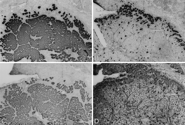
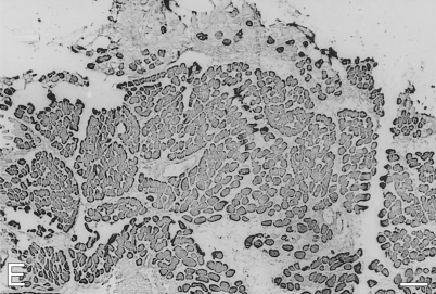
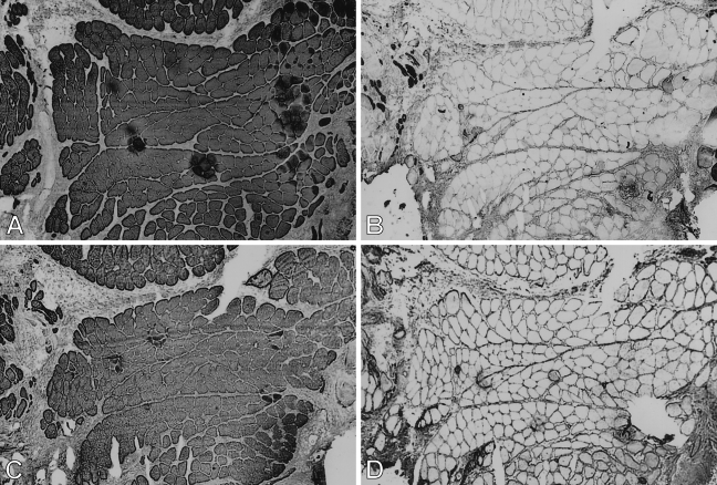
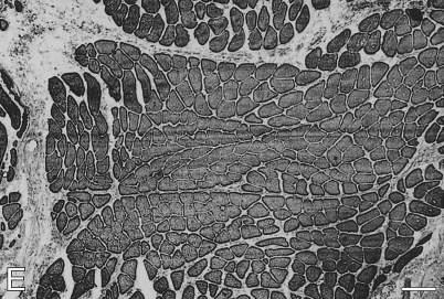
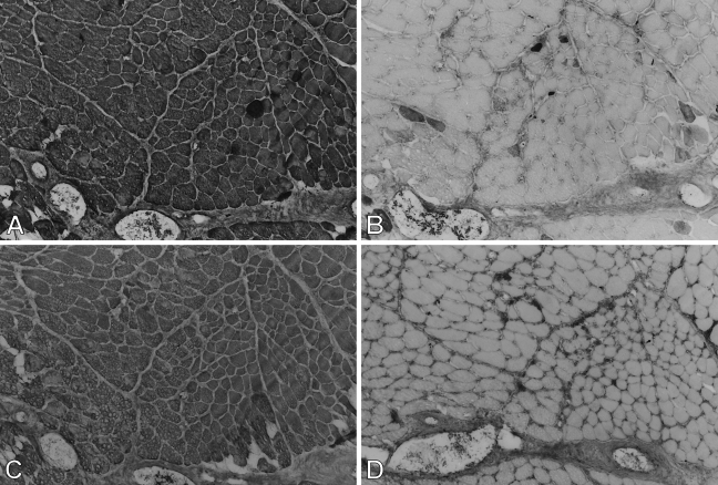
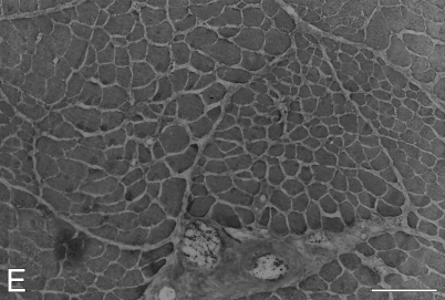
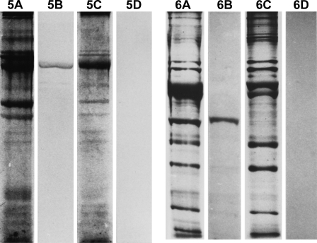
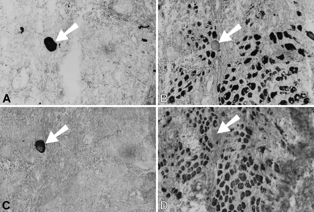
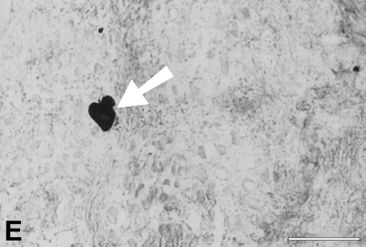
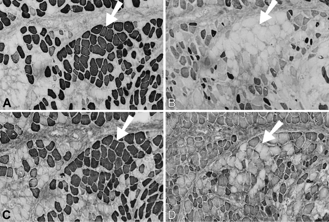
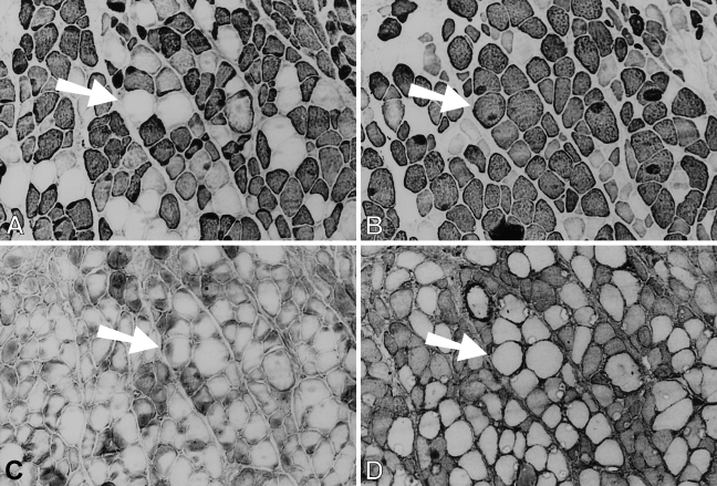
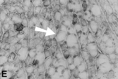
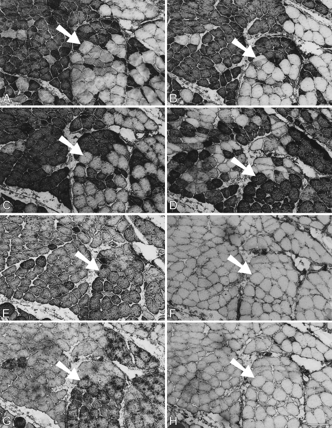
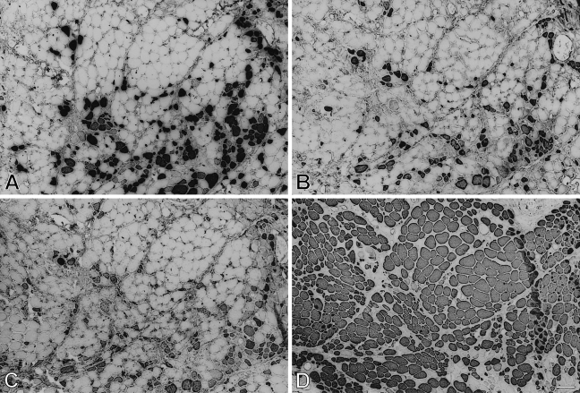
Cat jaw-closing muscles are a distinct muscle allotype characterized by the expression of masticatory-specific myofibrillar proteins. Transplantation studies showed that expression of masticatory myosin heavy chain (m-MyHC) is promoted by fast motor nerves, but suppressed by slow motor nerves. We investigated whether masticatory myosin-binding protein-C (m-MBP-C) and masticatory tropomyosin (m-Tm) are similarly regulated. Temporalis muscle strips were transplanted into limb muscle beds to allow innervation by fast or slow muscle nerve during regeneration. Regenerated muscles were examined postoperatively up to 168 days by peroxidase IHC using monoclonal antibodies to m-MyHC, m-MBP-C, and m-Tm. Regenerates in both muscle beds expressed fetal and slow MyHCs, m-MyHC, m-MBP-C, and m-Tm during the first 4 weeks. Longer-term regenerates innervated by fast nerve suppressed fetal and slow MyHCs, retaining m-MyHC, m-MBP-C, and m-Tm, whereas fibers innervated by slow nerve suppressed fetal MyHCs and the three masticatory-specific proteins, induced slow MyHC, and showed immunohistochemical characteristics of jaw-slow fibers. We concluded that expression of m-MBP-C and m-Tm is coregulated by m-MyHC and that neural impulses to limb slow muscle are capable of suppressing masticatory-specific proteins and to channel gene expression along the jaw-slow phenotype unique to jaw-closing muscle.


Figures

Figure 1
Figure 1
Low-power view of semiserial sections of temporalis muscle transplanted into the extensor digitorum longus (EDL) bed studied 9 days postoperatively, stained by immunoperoxidase with anti-masticatory myosin heavy chain monoclonal antibody (anti-m-MyHC MAb) 2F4 (A), anti-slow MyHC MAb NOQ-7-5-4D (B), anti-masticatory myosin-binding protein-C (anti-m-MBP-C) MAb 3F10 (C), anti-fetal MyHC antibody STE (D), and anti-masticatory-tropomyosin (anti-m-Tm) MAb 1H2 (E). Bar = 100 μm.
Figure 1
Figure 1
Low-power view of semiserial sections of temporalis muscle transplanted into the extensor digitorum longus (EDL) bed studied 9 days postoperatively, stained by immunoperoxidase with anti-masticatory myosin heavy chain monoclonal antibody (anti-m-MyHC MAb) 2F4 (A), anti-slow MyHC MAb NOQ-7-5-4D (B), anti-masticatory myosin-binding protein-C (anti-m-MBP-C) MAb 3F10 (C), anti-fetal MyHC antibody STE (D), and anti-masticatory-tropomyosin (anti-m-Tm) MAb 1H2 (E). Bar = 100 μm.
Figure 2
Figure 2
Immunoperoxidase staining of semiserial sections of temporalis muscle transplanted into the fast EDL bed examined 28 days postoperatively. Antibodies used were anti-m-MyHC (A), anti-slow MyHC (B), anti-m-MBP-C (C), anti-fetal MyHC (D), and anti-m-Tm (E). The arrow points to the same, relatively large diameter fiber in each panel that no longer expresses slow MyHC. Bar = 100 μm.
Figure 2
Figure 2
Immunoperoxidase staining of semiserial sections of temporalis muscle transplanted into the fast EDL bed examined 28 days postoperatively. Antibodies used were anti-m-MyHC (A), anti-slow MyHC (B), anti-m-MBP-C (C), anti-fetal MyHC (D), and anti-m-Tm (E). The arrow points to the same, relatively large diameter fiber in each panel that no longer expresses slow MyHC. Bar = 100 μm.
Figure 3
Figure 3
Low-power view of immunoperoxidase staining of semiserial sections of temporalis muscle transplanted into the EDL bed, studied 63 days postoperatively. Antibodies used were anti-m-MyHC (A), anti-slow MyHC (B), anti-m-MBP-C (C), anti-fetal MyHC (D), and anti-m-Tm (E). Bar = 100 μm.
Figure 3
Figure 3
Low-power view of immunoperoxidase staining of semiserial sections of temporalis muscle transplanted into the EDL bed, studied 63 days postoperatively. Antibodies used were anti-m-MyHC (A), anti-slow MyHC (B), anti-m-MBP-C (C), anti-fetal MyHC (D), and anti-m-Tm (E). Bar = 100 μm.
Figure 4
Figure 4
Immunoperoxidase staining of semiserial sections of temporalis muscle transplanted into the fast EDL bed studied 112 days postoperatively. Antibodies used were anti-m-MyHC (A), anti-slow MyHC (B), anti-m-MBP-C (C), anti-fetal MyHC (D), and anti-m-Tm (E). Bar = 50 μm.
Figure 4
Figure 4
Immunoperoxidase staining of semiserial sections of temporalis muscle transplanted into the fast EDL bed studied 112 days postoperatively. Antibodies used were anti-m-MyHC (A), anti-slow MyHC (B), anti-m-MBP-C (C), anti-fetal MyHC (D), and anti-m-Tm (E). Bar = 50 μm.
Figure 5
Figure 5
Analysis of myofibrillar proteins from 112-day regenerates of temporalis (A,B) and EDL (C,D) muscles innervated by the EDL nerve. A and C are protein-stained SDS-PAGE gels; B and D are Western blots using anti-m-MyHC.
Figure 6
Figure 6
Analysis of myofibrillar proteins from 112-day regenerates of temporalis (A,B) and EDL (C,D) muscles innervated by the EDL nerve. A and C are protein-stained SDS-PAGE gels; B and D are Western blots using anti-m-MBP-C.
Figure 7
Figure 7
High-power views of immunoperoxidase-stained semiserial sections of regenerating temporalis muscle 9 days after transplantation into the soleus muscle bed. Antibodies used were anti-m-MyHC (A), anti-slow MyHC (B), anti-m-MBP-C (C), anti-fetal MyHC (D), and anti-m-Tm (E). The arrow points to an island of necrotic tissue in each panel. Bar = 50 μm.
Figure 7
Figure 7
High-power views of immunoperoxidase-stained semiserial sections of regenerating temporalis muscle 9 days after transplantation into the soleus muscle bed. Antibodies used were anti-m-MyHC (A), anti-slow MyHC (B), anti-m-MBP-C (C), anti-fetal MyHC (D), and anti-m-Tm (E). The arrow points to an island of necrotic tissue in each panel. Bar = 50 μm.
Figure 8
Figure 8
Immunoperoxidase staining of semiserial sections of regenerating temporalis muscle 28 days after transplantation into the soleus muscle bed. Antibodies used were anti-m-MyHC (A), anti-slow MyHC (B), anti-m-MBP-C (C), anti-fetal MyHC (D), and anti-m-Tm (E). Arrow in each panel points to a group of large fibers that no longer expresses slow and fetal MyHCs. Bar = 50 μm.
Figure 8
Figure 8
Immunoperoxidase staining of semiserial sections of regenerating temporalis muscle 28 days after transplantation into the soleus muscle bed. Antibodies used were anti-m-MyHC (A), anti-slow MyHC (B), anti-m-MBP-C (C), anti-fetal MyHC (D), and anti-m-Tm (E). Arrow in each panel points to a group of large fibers that no longer expresses slow and fetal MyHCs. Bar = 50 μm.
Figure 9
Figure 9
Immunoperoxidase staining of semiserial sections of regenerating temporalis muscle 63 days after transplantation into the soleus muscle bed. Antibodies used were anti-m-MyHC (A), anti-slow MyHC (B), anti-m-MBP-C (C), anti-fetal MyHC (D), and anti-m-Tm (E). Arrow in each panel points to a pair of large fibers that have completely transformed into slow fibers. Bar = 50 μm.
Figure 9
Figure 9
Immunoperoxidase staining of semiserial sections of regenerating temporalis muscle 63 days after transplantation into the soleus muscle bed. Antibodies used were anti-m-MyHC (A), anti-slow MyHC (B), anti-m-MBP-C (C), anti-fetal MyHC (D), and anti-m-Tm (E). Arrow in each panel points to a pair of large fibers that have completely transformed into slow fibers. Bar = 50 μm.
Figure 10
Figure 10
Immunoperoxidase staining of semiserial sections of regenerating temporalis muscle 91 days after transplantation into the soleus muscle bed. Antibodies used were anti-m-Tm (A), anti-m-MBP-C (B), anti-m-MyHC (C), anti-slow MyHC (D), MAb 1A10 (E), anti-fast MyHC (F), anti-limb Tm CH1 (G), and anti-fetal MyHC (H). The arrow in each panel points to a jaw-slow fiber. Bar = 100 μm.
Figure 11
Figure 11
Immunoperoxidase staining of semiserial sections of temporalis muscle regenerated in the soleus bed 168 days postoperatively. Antibodies used were anti-m-MyHC (A), anti-m-Tm (B), anti-m-MBP-C (C), and anti-slow MyHC (D). Bar = 100 μm.

References

    1. Adams GR, McCue SA, Zeng M, Baldwin KM (1999) Time course of myosin heavy chain transitions in neonatal rats: importance of innervation and thyroid state. Am J Physiol 276:R954–961 - PubMed
    1. Allen DL, Sartorius CA, Sycuro LK, Leinwand LA (2001) Different pathways regulate expression of the skeletal myosin heavy chain genes. J Biol Chem 276:43524–43533 - PubMed
    1. Bar A, Pette D (1988) Three fast myosin heavy chains in adult rat skeletal muscle. FEBS Lett 235:153–155 - PubMed
    1. Bottinelli R, Schiaffino S, Reggiani C (1991) Force-velocity relations and myosin heavy chain isoform compositions of skinned fibres from rat skeletal muscle. J Physiol 437:655–672 - PMC - PubMed
    1. Chin ER, Olson EN, Richardson JA, Yang Q, Humphries C, Shelton JM, Wu H, et al. (1998) A calcineurin-dependent transcriptional pathway controls skeletal muscle fiber type. Genes Dev 12:2499–2509 - PMC - PubMed

Publication types