

Metastasis in an orthotopic murine model of melanoma is independent of RAS/RAF mutation
- PMID: 20679910
- PMCID: PMC2972650
- DOI: 10.1097/CMR.0b013e328336ee17
Metastasis in an orthotopic murine model of melanoma is independent of RAS/RAF mutation
Abstract
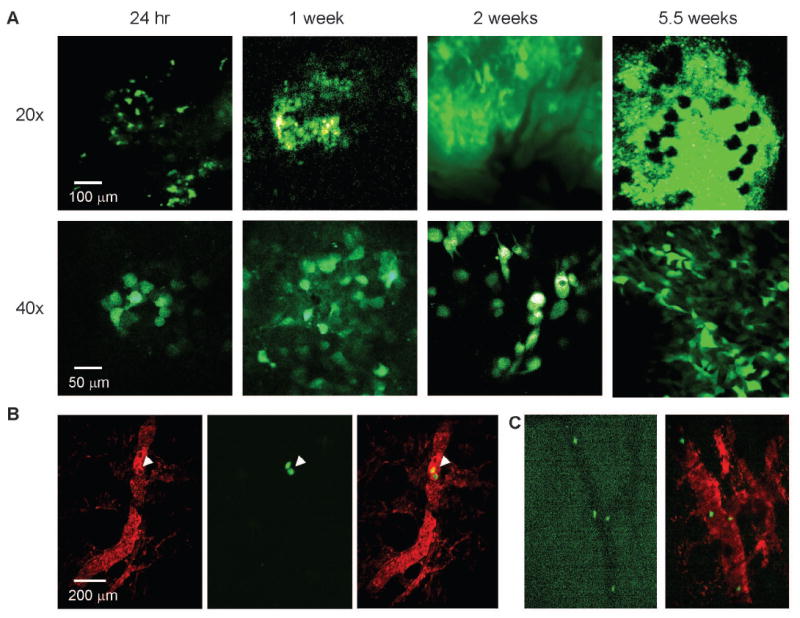
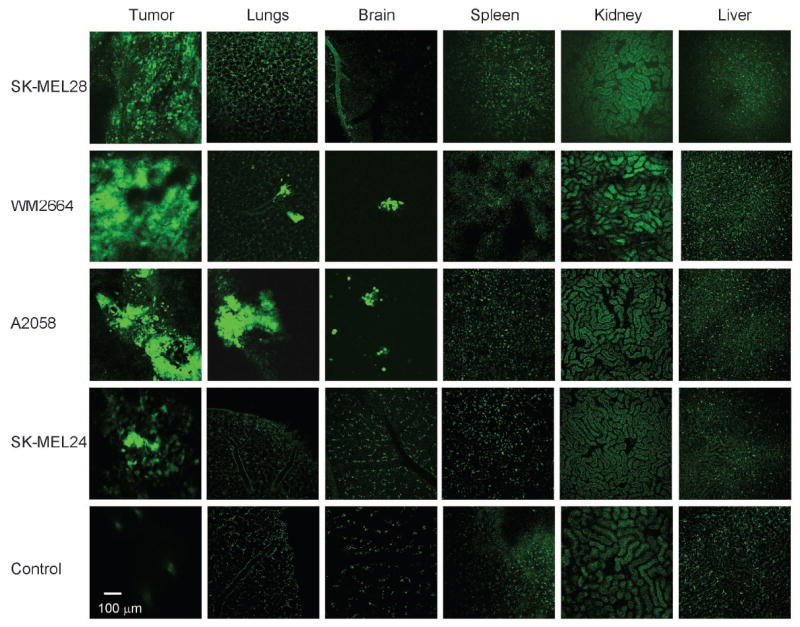
Melanoma is the most lethal skin tumor in large part because of a propensity for early metastasis. Good models of this most clinically relevant feature of melanoma are lacking. Here, we report the development of an in-vivo model of metastasis that relies on orthotopic injection of green fluorescent protein-tagged lines in immunodeficient mice, serial intravital imaging of tumor progression, and quantification of distant spread by two-photon laser scanning microscopy, immunohistochemistry, and real-time PCR analysis. Using this system, we report an assessment of the in-vivo growth and metastatic properties of 11 well-characterized human melanoma cell lines. A subset of lines showed rapid in-vivo growth with invasion of host vasculature and distant seeding of viscera in this system. The ability to form metastasis in vivo did not correlate with three-dimensional collagen invasion in vitro. Surprisingly, similar lines in terms of molecular genetic events differed markedly in their propensity to metastasize to distant organs such as brain and lung. In particular, two lines harboring B-RAF mutation and high levels of phosphorylated ERK and AKT were reproducibly unable to form tumors after orthotopic injection. Similarly, two previously identified RAS/RAF wildtype 'epithelial like' lines that do not have elevated phosphorylated ERK and AKT or express TWIST1 mRNA still showed a pronounced ability for orthotopic growth and metastatic spread. All the metastatic cell lines in this model showed increased NEDD9 expression, but NEDD9 lentiviral overexpression did not convey a metastatic phenotype on nonmetastatic cells. These data suggest that melanoma metastasis is a molecularly heterogeneous process that may not require epithelial-to-mesenchymal transition or ERK activation, although both may facilitate the process.
Figures


Similar articles
-
The BRAF(V600E) inhibitor, PLX4032, increases type I collagen synthesis in melanoma cells.Matrix Biol. 2015 Oct;48:66-77. doi: 10.1016/j.matbio.2015.05.007. Epub 2015 May 16. Matrix Biol. 2015. PMID: 25989506 Free PMC article.
-
Lack of extracellular signal-regulated kinase mitogen-activated protein kinase signaling shows a new type of melanoma.Cancer Res. 2007 Feb 15;67(4):1502-12. doi: 10.1158/0008-5472.CAN-06-3311. Cancer Res. 2007. PMID: 17308088
-
S100A4 expression with reduced E-cadherin expression predicts distant metastasis of human malignant melanoma cell lines in the NOD/SCID/gammaCnull (NOG) mouse model.Oncol Rep. 2005 Sep;14(3):633-7. Oncol Rep. 2005. PMID: 16077966
-
Roads to melanoma: Key pathways and emerging players in melanoma progression and oncogenic signaling.Biochim Biophys Acta. 2016 Apr;1863(4):770-84. doi: 10.1016/j.bbamcr.2016.01.025. Epub 2016 Feb 1. Biochim Biophys Acta. 2016. PMID: 26844774 Review.
-
Ras, Raf, and MAP kinase in melanoma.Adv Anat Pathol. 2013 Jul;20(4):217-26. doi: 10.1097/PAP.0b013e3182976c94. Adv Anat Pathol. 2013. PMID: 23752084 Review.
Cited by
-
Prioritizing therapeutic targets using patient-derived xenograft models.Biochim Biophys Acta. 2015 Apr;1855(2):223-34. doi: 10.1016/j.bbcan.2015.03.002. Epub 2015 Mar 14. Biochim Biophys Acta. 2015. PMID: 25783201 Free PMC article. Review.
-
Orthotopic and metastatic tumour models in preclinical cancer research.Pharmacol Ther. 2024 May;257:108631. doi: 10.1016/j.pharmthera.2024.108631. Epub 2024 Mar 11. Pharmacol Ther. 2024. PMID: 38467308 Free PMC article. Review.
-
Preclinical Solid Tumor Models to Study Novel Therapeutics in Brain Metastases.Curr Protoc. 2021 Nov;1(11):e284. doi: 10.1002/cpz1.284. Curr Protoc. 2021. PMID: 34762346 Free PMC article.
-
Imaging the pharmacology of nanomaterials by intravital microscopy: Toward understanding their biological behavior.Adv Drug Deliv Rev. 2017 Apr;113:61-86. doi: 10.1016/j.addr.2016.05.023. Epub 2016 Jun 4. Adv Drug Deliv Rev. 2017. PMID: 27266447 Free PMC article. Review.
-
Intravital imaging of a spheroid-based orthotopic model of melanoma in the mouse ear skin.Intravital. 2013;2(2):e25805. doi: 10.4161/intv.25805. Epub 2013 Apr 1. Intravital. 2013. PMID: 28748125 Free PMC article.
References
-
- Anbari KK, Schuchter LM, Bucky LP, Mick R, Synnestvedt M, Guerry D, et al. Melanoma of unknown primary site: presentation, treatment, and prognosis--a single institution study. University of Pennsylvania Pigmented Lesion Study Group. Cancer. 1997;79:1816–21. - PubMed
-
- Baab GH, McBride CM. Malignant melanoma: the patient with an unknown site of primary origin. Arch Surg. 1975;110:896–900. - PubMed
-
- Chang P, Knapper WH. Metastatic melanoma of unknown primary. Cancer. 1982;49:1106–11. - PubMed
-
- Gattoni-Celli S, Byers SH, Calorini L, Ferrone S. Organ-Specific Metastases in Melanoma: Experimental Animal Models. Pigment Cell Res. 1993;6:981–384. - PubMed
Publication types
MeSH terms
Substances
Grants and funding
LinkOut - more resources
Full Text Sources
Other Literature Sources
Medical
Research Materials
Miscellaneous
