NGF and NGF-receptor expression of cultured immortalized human corneal endothelial cells
- PMID: 20680101
- PMCID: PMC2913141
NGF and NGF-receptor expression of cultured immortalized human corneal endothelial cells
Abstract
Purpose: Several growth factors, including nerve growth factor (NGF) and vascular endothelial growth factor (VEGF), play an important role in the homeostasis of the ocular surface. The involvement of both these growth factors in the pathophysiology of intraocular tissues has been extensively investigated. Despite the expression of NGF receptors by corneal endothelium, to date the role of NGF on the endothelial cell remains to be determined. Using a clonal cell line of human corneal endothelial cells, the aim of this study was to investigate the expression of the NGF-receptor and the potential partnership of NGF and VEGF in maintaining cell viability in vitro.
Methods: A human endothelial cell line (B4G12), was cultured under serum-free conditions as previously described with and without addition of different concentrations of NGF, anti-NGF-antibody (ANA), or VEGF for 4 days and these cells were used for immuno-istochemical, biochemical, and molecular analyses.
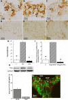
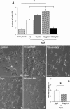
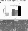
Results: NGF induces overexpression of NGF-receptors and synthesis and release of VEGF by endothelial cells and these cells are able to produce and secrete NGF.
Conclusions: These observations indicate that human corneal endothelial cells are receptive to the action of NGF and that these cells may regulate NGF activity through autocrine/paracrine mechanisms.
Figures


Similar articles
-
Nerve growth factor-induced migration of endothelial cells.J Pharmacol Exp Ther. 2005 Dec;315(3):1220-7. doi: 10.1124/jpet.105.093252. Epub 2005 Aug 25. J Pharmacol Exp Ther. 2005. PMID: 16123305
-
Nerve growth factor is critical requirement for in vitro angiogenesis in gastric endothelial cells.Am J Physiol Gastrointest Liver Physiol. 2016 Nov 1;311(5):G981-G987. doi: 10.1152/ajpgi.00334.2016. Epub 2016 Oct 13. Am J Physiol Gastrointest Liver Physiol. 2016. PMID: 27742705
-
In vivo antivascular endothelial growth factor treatment induces corneal endothelium apoptosis in rabbits through changes in p75NTR-proNGF pathway.J Cell Physiol. 2018 Nov;233(11):8874-8883. doi: 10.1002/jcp.26806. Epub 2018 Jun 1. J Cell Physiol. 2018. PMID: 29856479
-
[Cardiovascular effects of nerve growth factor analytical review. Part 2].Fiziol Cheloveka. 2011 May-Jun;37(3):109-28. Fiziol Cheloveka. 2011. PMID: 21780688 Review. Russian.
-
Unraveling the story of NGF-mediated sensitization of nociceptive sensory neurons: ON or OFF the Trks?Mol Interv. 2007 Feb;7(1):26-41. doi: 10.1124/mi.7.1.6. Mol Interv. 2007. PMID: 17339604 Review.
Cited by
-
Evaluation of moxifloxacin-induced cytotoxicity on human corneal endothelial cells.Sci Rep. 2021 Mar 18;11(1):6250. doi: 10.1038/s41598-021-85834-x. Sci Rep. 2021. PMID: 33737688 Free PMC article.
-
An In Vitro Model of the Blood-Brain Barrier for the Investigation and Isolation of the Key Drivers of Barriergenesis.Adv Healthc Mater. 2024 Dec;13(32):e2303777. doi: 10.1002/adhm.202303777. Epub 2024 Aug 5. Adv Healthc Mater. 2024. PMID: 39101628 Free PMC article.
-
The Role of Nerve Growth Factor in Maintaining Proliferative Capacity, Colony-Forming Efficiency, and the Limbal Stem Cell Phenotype.Stem Cells. 2019 Jan;37(1):139-149. doi: 10.1002/stem.2921. Stem Cells. 2019. PMID: 30599086 Free PMC article.
-
Tear Neuromediators in Subjects with and without Dry Eye According to Ocular Sensitivity.Chonnam Med J. 2022 Jan;58(1):37-42. doi: 10.4068/cmj.2022.58.1.37. Epub 2022 Jan 25. Chonnam Med J. 2022. PMID: 35169558 Free PMC article.
-
Expression of Hormones' Receptors in Human Corneal Endothelium from Fuchs' Dystrophy: A Possible Gender' Association.J Clin Med. 2024 Jun 27;13(13):3787. doi: 10.3390/jcm13133787. J Clin Med. 2024. PMID: 38999352 Free PMC article.
References
-
- Levi-Montalcini R. The nerve growth factor 35 years later. Science. 1987;237:1154–62. - PubMed
-
- Aloe L and Calza L. editors. NGF and Related Molecules in Health and Disease. Progr Brain Res Elsevier 2004.
-
- Oku H, Ikeda T, Honma Y, Sotozono C, Nishida K, Nakamura Y, Kida T, Kinoshita S. Gene expression of neurotrophins and their high-affinity Trk receptors in cultured human Müller cells. Ophthalmic Res. 2002;34:38–42. - PubMed
-
- Siliprandi R, Canella R, Carmignoto G. Nerve growth factor promotes functional recovery of retinal ganglion cells after ischemia. Invest Ophthalmol Vis Sci. 1993;34:3232–45. - PubMed
Publication types
MeSH terms
Substances
LinkOut - more resources
Full Text Sources
Other Literature Sources
