Size of third and fourth ventricle in obstructive and communicating acute hydrocephalus after aneurysmal subarachnoid hemorrhage
- PMID: 20680324
- PMCID: PMC3016153
- DOI: 10.1007/s00415-010-5678-1
Size of third and fourth ventricle in obstructive and communicating acute hydrocephalus after aneurysmal subarachnoid hemorrhage
Abstract
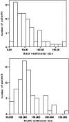
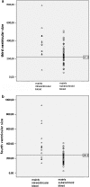
In patients with acute hydrocephalus after aneurysmal subarachnoid haemorrhage (SAH), lumbar drainage is possible if the obstruction is in the subarachnoid space (communicating hydrocephalus). In case of intraventricular obstruction (obstructive hydrocephalus), ventricular drainage is the only option. A small fourth ventricle is often considered a sign of obstructive hydrocephalus. We investigated whether the absolute or relative size of the fourth ventricle can indeed distinguish between these two types of hydrocephalus. On CT-scans of 76 consecutive patients with acute headache but normal CT and CSF, we measured the cross-sectional surface of the third and fourth ventricle to obtain normal planimetric values. Subsequently we performed the same measurements on 117 consecutive SAH patients with acute hydrocephalus. These patients were divided according to the distribution of blood on CT-scan into three groups: mainly intraventricular blood (n=15), mainly subarachnoid blood (n=54) and both intraventricular and subarachnoid blood (n=48). The size of the fourth ventricle exceeded the upper limit of normal in 2 of the 6 (33%) patients with intraventricular blood but without haematocephalus, and in 15 of the 54 (28%) patients with mainly subarachnoid blood. The mean ratio between the third and fourth ventricle was 1.45 (SD 0.66) in patients with intraventricular blood and 1.42 (SD 0.91) in those with mainly subarachnoid blood. Neither fourth ventricular size nor the ratio between the third and fourth ventricles discriminates between the two groups. A small fourth ventricle does not necessarily accompany obstructive hydrocephalus and is therefore not a contraindication for lumbar drainage.
Figures


Similar articles
-
The Volume of the Third Ventricle as a Prognostic Marker for Shunt Dependency After Aneurysmal Subarachnoid Hemorrhage.World Neurosurg. 2017 Dec;108:107-111. doi: 10.1016/j.wneu.2017.08.129. Epub 2017 Sep 1. World Neurosurg. 2017. PMID: 28867328
-
Factors related to acute hydrocephalus after subarachnoid hemorrhage.Acta Radiol. 2004 May;45(3):333-9. doi: 10.1080/02841850410004274. Acta Radiol. 2004. PMID: 15239431
-
Predictors of long-term shunt-dependent hydrocephalus after aneurysmal subarachnoid hemorrhage. Clinical article.J Neurosurg. 2010 Oct;113(4):774-80. doi: 10.3171/2010.2.JNS09376. J Neurosurg. 2010. PMID: 20367072
-
Acute hydrocephalus following post-traumatic peri-mesencephalic subarachnoid hemorrhage: an uncommon and potentially fatal event in children.Childs Nerv Syst. 2023 Mar;39(3):577-581. doi: 10.1007/s00381-023-05842-2. Epub 2023 Jan 13. Childs Nerv Syst. 2023. PMID: 36637469 Review.
-
Hydrocephalus after aneurysmal subarachnoid hemorrhage.Neurosurg Clin N Am. 2010 Apr;21(2):263-70. doi: 10.1016/j.nec.2009.10.013. Neurosurg Clin N Am. 2010. PMID: 20380968 Review.
Cited by
-
Operative planning aid for optimal endoscopic third ventriculostomy entry points in pediatric cases.Childs Nerv Syst. 2017 Feb;33(2):269-273. doi: 10.1007/s00381-016-3320-y. Epub 2017 Jan 18. Childs Nerv Syst. 2017. PMID: 28101675 Free PMC article.
-
Progress in cerebrovascular disease research in the last year.J Neurol. 2012 Feb;259(2):391-4. doi: 10.1007/s00415-012-6408-7. Epub 2012 Jan 19. J Neurol. 2012. PMID: 22258481 Review.
References
MeSH terms
LinkOut - more resources
Full Text Sources
Medical

