. 2010 Sep;28(9):1560-7.
doi: 10.1002/stem.491.

Loss of Bright/ARID3a function promotes developmental plasticity

Affiliations

Loss of Bright/ARID3a function promotes developmental plasticity

Guangyu An et al. Stem Cells. 2010 Sep.

Abstract

B-cell regulator of immunoglobulin heavy chain transcription (Bright)/ARID3a, an A+T-rich interaction domain protein, was originally discovered in B lymphocyte lineage cells. However, expression patterns and high lethality levels in knockout mice suggested that it had additional functions. Three independent lines of evidence show that functional inhibition of Bright results in increased developmental plasticity. Bright-deficient cells from two mouse models expressed a number of pluripotency-associated gene products, expanded indefinitely, and spontaneously differentiated into cells of multiple lineages. Furthermore, direct knockdown of human Bright resulted in colonies capable of expressing multiple lineage markers. These data suggest that repression of this single molecule confers adult somatic cells with new developmental options.


Conflict of interest statement

Disclosure of potential conflicts of interest: The authors indicate no potential conflicts of interest.

Figures

Fig. 1
Fig. 1
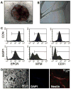
Bright deficient cultures spontaneously differentiate into multiple lineages. (A) A representative embryoid-like body from Bright−/− spleen. (B) Bright−/− spleen cells grown in DMEM with 10% FCS and endothelial cell growth factors form multicellular, tube-like structures. (4× magnification) (C) Flow cytometry of a control (CON) mouse hemangioma cell line (gift of C. Esmon, this institution) and Bright−/− cultures (BrSP1) reveals expression of the endothelial protein C receptor (EPCR), mouse thrombomodulin (MTM), and CD31 endothelial markers. (D) Bright−/− bone marrow cultures seeded into matrigel developed branched neuron-like cell clusters after three weeks (left, 20x magnification). Cells were stained with DAPI to identify nuclei (middle) or anti-nestin (right), a neuronal marker.
Fig. 2
Fig. 2
Bright-deficient cultures express pluripotency-associated markers. (A) RT-PCR assays were performed with normal spleen (WT1), 2 Bright−/− whole spleen cultures (BrSPS1 and BrSPS2, cultured 6 months) and conventional ES cells. (B) Cloned Bright−/− kidney cells (BrKPS5) formed colonies on MEFs, with small nuclei (DAPI) and expressed the early stem cell marker, SSEA-1. The MEF monolayer from the same culture did not stain with SSEA-1, nor did any of the cells stain with the isotype-matched control (Iso).
Fig. 3
Fig. 3
DN Bright cultures express Nanog and appear to be B lymphocyte-derived. (A) DN Bright whole spleen cultures (DN1 and DN2), control spleen cultures (WT1) and ES cells were assessed for gene expression by RT-PCR. (B) Genomic DNA from fresh spleen cells (spleen), a representative DN Bright cultured cells (DN1, in culture 8 months) and negative control ES and MEF cells (Con1 and 2, respectively) was amplified using primers for the indicated JH rearrangements (arrows). (C) Sorted pre-B cells from DN-Bright mice formed granular colonies after 3 weeks (left two panels), while control pre-B cells maintained their original morphology (right panel).
Fig. 4
Fig. 4
ARID3a knockdown in a human cell line induces alterations in colony morphology. (A) Western blotting indicates that ARID3a is abundantly expressed in human epithelial kidney 293T cells as compared to the human fibroblast cell lines WL-38, BJ-h, BJ and IM-R90. Actin levels show relative loading. Lane 2 is empty. (B) RT-PCR of ARID3a RNA levels demonstrates efficient knockdown in 2 ARID3a-inhibited clones (Bri1 and 2) compared to control 293T cells. GAPDH served as a loading control. (C) ARID3a-inhibited 293T cells (BriPS) exhibited increasing ES cell-like colony morphology compared to control 293T cultures with additional passages (P7 versus P10).
Fig. 5
Fig. 5
ARID3a knockdown in human 293T cells induces expression of pluripotency associated markers. (A) QRT-PCR of two ARID3a-inhibited clones versus the 293T parental cells shows fold-induction levels of KLf4, Oct4, Sox2 and c-myc transcripts relative to GAPDH. (B) Nuclear (DAPI) and immunohistochemical staining (Oct4 and Nanog) of two ARID3a deficient clones, Bri1 and Bri11, along with iostype staining controls (Iso), right two panels.
Fig. 6
Fig. 6
Human Bright deficient clones generated by ARID3a knockdown differentiate in vitro to express markers for all three germ line lineages. (A) Conventional mouse ES cells (top panels), Bri1 (middle panels) and parental 293T cells (bottom panels) were stained for α-fetoprotein (AFP), smooth muscle actin (SMA), β-3-tubulin (BIIIT) or an isotype control (ISO). (B) DAPI nuclear stains are shown for all panels in (A). (C) ARID3a-deficient cells did not form teratomas in Nod/Scid mice. Shown are representative sections of early neoplastic cells formed from Bri1 (left) and a teratoma from conventional ES cells (right). Magnification is 40×.

References

    1. Webb CF, Das C, Eaton S, et al. Novel protein-DNA interactions associated with increased immunoglobulin transcription in response to antigen plus interleukin-5. Mol Cell Biol. 1991;11:5197–5205. - PMC - PubMed
    1. Herrscher RF, Kaplan MH, Lelsz DL, et al. The immunoglobulin heavy-chain matrix-associating regions are bound by Bright: a B cell-specific trans-activator that describes a new DNA-binding protein family. Genes Dev. 1995;9:3067–3082. - PubMed
    1. Wilsker D, Patsialou A, Dallas PB, et al. ARID proteins: a diverse family of DNA binding proteins implicated in the control of cell growth, differentiation, and development. Cell Growth Differ. 2002;13:95–106. - PubMed
    1. Wilsker D, Probst L, Wain HM, et al. Nomenclature of the ARID family of DNA-binding proteins. Genomics. 2005;86:242–251. - PubMed
    1. Peeper DS, Shvarts A, Brummelkamp T, et al. A functional screen identifies hDRIL1 as an oncogene that rescues RAS- induced senescence. Nat Cell Biol. 2002;4 :148–153. - PubMed

Publication types

MeSH terms