. 2010 Sep 3;9(9):4433-42.
doi: 10.1021/pr100662s.

Protein kinase-inhibitor database: structural variability of and inhibitor interactions with the protein kinase P-loop

Affiliations

Protein kinase-inhibitor database: structural variability of and inhibitor interactions with the protein kinase P-loop

Ronak Y Patel et al. J Proteome Res. .

Abstract

Structure-based drug design of protein-kinase inhibitors has been facilitated by availability of an enormous number of structures in the Protein Databank (PDB), systematic analyses of which can provide insight into the factors that govern ligand-protein kinase interactions and into the conformational variability of the protein kinases. In this study, a nonredundant database containing 755 unique, curated, and annotated PDB protein kinase-inhibitor complexes (each consisting of a single protein kinase chain, a ligand, and water molecules around the ligand) was created. With this dataset, analyses were performed of protein conformational variability and interactions of ligands with 11 P-loop residues. Analysis of ligand-protein interactions included ligand atom preference, ligand-protein hydrogen bonds, and the number and position of crystallographic water molecules around important P-loop residues. Analysis of variability in the conformation of the P-loop considered backbone and side-chain dihedral angles, and solvent accessible surface area (SASA). A distorted conformation of the P-loop was observed for some of the protein kinase structures. Lower SASA was observed for the hydrophobic residue in beta1 of several members of the AGC family of protein kinases. Our systematic studies were performed amino acid-by-amino acid, which is unusual for analyses of protein kinase-inhibitor complexes.


Figures

Figure 1
Figure 1
(A) Typical location of the P-loop in a representative protein kinase structure (1A9U) is shown as anti-parallel magenta strands β1 and β2 connected by a magenta loop. Secondary structure elements which interact with ATP-competitive inhibitors are labeled. (B) The P-loop is magnified and individual Cα atoms are shown as magenta spheres. The residue nomenclature is shown, with positive and negative numbering relative to G0. Side-chains are shown as sticks. (C) The locations of water molecules (oxygen atoms) within 3 Å of the P-loop residues L–3 (orange spheres), G–2 (blue spheres) and V5 (cyan spheres) are shown for all 532 structures. The amino-acid backbone of each structure was superimposed on the backbone of the corresponding amino-acid of structure 1A9U. Ligand SB2 (co-crystallized in 1A9U) is shown with green sticks. The protein backbone and side chains of 1A9U are shown as magenta sticks. The side-chains of residues L–3 and V5 of other protein structures are shown as thin magenta sticks.
Figure 2
Figure 2
Number of structures containing at least one kinase domain submitted to RCSB Protein Data Bank (PDB) per year. The date of submission was derived from the PDB file HEADER records.
Figure 3
Figure 3
(A). Residue conservation in the P-loop represented using sequence logos. The sequence logos were created using the sequences of 532 structures containing a complete P-loop in terms of backbone and side-chain atoms. The Y-axis for both panels is the residue position from panel A, which is determined relative to the central glycine position in the P-loop (G0). (B) Conservation of secondary structure, represented using sequence logos. The secondary structure was computed using Stride, where E, C and T represent the secondary structure type of the amino-acid as extended configuration of a β-sheet, coil, and turn, respectively.
Figure 4
Figure 4
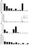
Interactions of the P-loop residues (L–3 to X7) of protein kinases with ligands. (A) Number of protein structures having at least one non-water atom within 3 Å of amino-acid residues. (B) Number of various atoms (carbon, C; nitrogen, N; oxygen, O; and halogens, F, Cl, Br and I) within 3 Å of protein residue. (C) Number of ligand–protein hydrogen bonds as a function of protein residue. (D) Number of structures with at least one water molecules within 3 Å of protein residue.
Figure 5
Figure 5
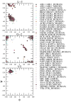
Backbone dihedral angles, φ and ψ (in °), computed using stride for the three residues (L–3, G–2 and V5) which make contact with inhibitors in the most structures in the dataset. Data is shown only for 485 proteins with >2 structures in the original dataset of 755 structures.
Figure 6
Figure 6
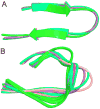
(A) Usually observed fully-extended conformation (PDBs: ABL1/ABL1_HUMAN, 2hz0; JNK3/MK10_HUMAN, 1pmn; p38α/MK14_HUMAN, 1w82) and (B) Rarer distorted conformation of the P-loop, rendered as ribbons (PDBs: ABL1/ABL1_HUMAN: 2hzi, 2f4j, 1opl, 3cs9, 2hiw, 2e2b, 2hyy; ABL1/ABL1_MOUSE: 1m52, 1opk, 3dk3, 2qoh, 1iep and 1fpu; p38α/MK14_HUMAN: 1w84, 2i0h, 3d7z, 1w7h, 3bx5, 1wbw, 1zzl, 3e92, 3c5u, 1yqj, 3ctq; ABL2/ABL2_HUMAN: 3gvu and 3hmi; AurA/STK6_MOUSE: 3dj5 and 3dj6; FGFR1/FGFR1_HUMAN: 3c4f; CK2α1/CSK21_HUMAN: 2zjw; JNK3/MK10_HUMAN: 2r9s). The following color coding was used for individual proteins: green, ABL1; blue purple, JNK3; spring green, p38α; blue, ABL2; orange, AurA; magenta, FGFR1 and pink, CK2α1. This snapshot was created using the maestro module of the Schrödinger software suite.
Figure 7
Figure 7
Conformational flexibility of amino-acid side chains represented by side chain dihedral angles χ1, χ2 and χ3 for two residues (A) L–3 and (B) V5. The side chain dihedral angles were calculated using the dangle program.
Figure 8
Figure 8
Solvent accessible surface area (SASA) calculated using NAccess for the different protein kinase groups for three important P-loop residues making significant contacts with inhibitors (L–3, G–2 and V5). The single SASA value plotted for each protein represents the average over all the structures in the database for that particular protein; the error bars show the standard deviation in SASA for all the structures for that protein.
Figure 9
Figure 9
Representative example of C-terminal extension (rendered as black ribbons) of AGC-family members, 2c1b, PKaCA (left panel); 2jdo, AKT2 (middle panel) and 3d9v, ROCK1 (right panel) interacting with the L–3 residue (rendered as magenta spheres) of the P-loop. The P-loop is rendered as orange ribbons. This interaction is responsible for reduction in solvent accessible surface area of residue L–2 for the crystal structures of these proteins.

Similar articles

Cited by

References

    1. Manning G, Whyte DB, Martinez R, Hunter T, Sudarsanam S. The protein kinase complement of the human genome. Science. 2002;298(5600):1912–34. - PubMed
    1. Eswaran J, Knapp S. Insights into protein kinase regulation and inhibition by large scale structural comparison. Biochim Biophys Acta. 2009;1804(3):429–432. - PMC - PubMed
    1. Cherry M, Williams DH. Recent kinase and kinase inhibitor X-ray structures: mechanisms of inhibition and selectivity insights. Curr Med Chem. 2004;11(6):663–73. - PubMed
    1. Rubin GM, Yandell MD, Wortman JR, Gabor Miklos GL, Nelson CR, Hariharan IK, Fortini ME, Li PW, Apweiler R, Fleischmann W, Cherry JM, Henikoff S, Skupski MP, Misra S, Ashburner M, Birney E, Boguski MS, Brody T, Brokstein P, Celniker SE, Chervitz SA, Coates D, Cravchik A, Gabrielian A, Galle RF, Gelbart WM, George RA, Goldstein LS, Gong F, Guan P, Harris NL, Hay BA, Hoskins RA, Li J, Li Z, Hynes RO, Jones SJ, Kuehl PM, Lemaitre B, Littleton JT, Morrison DK, Mungall C, O’Farrell PH, Pickeral OK, Shue C, Vosshall LB, Zhang J, Zhao Q, Zheng XH, Lewis S. Comparative genomics of the eukaryotes. Science. 2000;287(5461):2204–15. - PMC - PubMed
    1. Huse M, Kuriyan J. The conformational plasticity of protein kinases. Cell. 2002;109(3):275–82. - PubMed

Publication types