. 2010 Sep 1;82(17):7218-26.
doi: 10.1021/ac100989q.

Characterization of oligodeoxynucleotides and modifications by 193 nm photodissociation and electron photodetachment dissociation

Affiliations

Characterization of oligodeoxynucleotides and modifications by 193 nm photodissociation and electron photodetachment dissociation

Suncerae I Smith et al. Anal Chem. .

Abstract

Ultraviolet photodissociation (UVPD) at 193 nm is compared to collision induced dissociation (CID) for sequencing and determination of modifications of multideprotonated 6-20-mer oligodeoxynucleotides. UVPD at 193 nm causes efficient charge reduction of the deprotonated oligodeoxynucleotides via electron detachment, in addition to extensive backbone cleavages to yield sequence ions of relatively low abundance, including w, x, y, z, a, a-B, b, c, and d ions. Although internal ions populate UVPD spectra, base loss ions from the precursor are absent. Subsequent CID of the charge-reduced oligodeoxynucleotides formed upon electron detachment, in a net process called electron photodetachment dissociation (EPD), results in abundant sequence ions in terms of w, z, a, a-B, and d products, with a marked decrease in the abundance of precursor base loss ions and internal fragments. Complete sequencing was possible for virtually all oligodeoxynucleotides studied. EPD of three modified oligodeoxynucleotides, a methylated oligodeoxynucleotide, a phosphorothioate-modified oligodeoxynucleotide, and an ethylated-oligodeoxynucleotide, resulted in specific and extensive backbone cleavages, specifically, w, z, a, a-B, and d products, which allowed the modification site(s) to be pinpointed to a more specific location than by conventional CID.


Figures

Figure 1
Figure 1
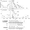
MS/MS spectra of ss12 by (a) UVPD of 4- (one UV pulse, 5mJ), (b) EPD of 3-• (one UV pulse, 5mJ; then 28% normalized collision energy, 30 ms), and (c) CID 4- (20% normalized collision energy, 30 ms), and (d) IRMPD 4- (5.0 ms irradiation, 10 W). Precursor ions are noted with a star.
Figure 2
Figure 2
Electron detachment yield for the 2- and 3- charge states of dA6, dG6, dC6, and dT6 upon one UV laser pulse (5 mJ). The fraction of charge-reduced species was determined based on the abundances of all radical products and the charge- reduced precursor, with the assumption that all radical product ions are derived from decomposition of the charge reduced precursor. The standard deviation was calculated from three experiments.
Figure 3
Figure 3
Number of diagnostic sequence ions [a-B and w for CID; a, a-B, d, w, z, and other ions (including b, c, x, y) for UVPD; a, a-B, d, w and z for EPD] are displayed in bar graph form for each observed charge state for ss8, ss12, ss16, and ss20.
Figure 4
Figure 4
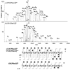
MS/MS spectra of Mss10 by (a) EPD of 3-• (one UV pulse, 5mJ; then 8% normalized collision energy, 30 ms), and (b) CID 3- (8% normalized collision energy, 30 ms). Precursor ions are noted with a star. Bolded and italicized product ions retain the modification. A summary of the product ions formed from EPD and CID for the methylated single strand 10-mer (c), in which the slash marks represent cleavages at that location. A crescent above the slash mark represents a product containing the methyl modification. .
Figure 5
Figure 5
MS/MS spectra of PSss12 by (a) EPD of 4-• (one UV pulse, 5 mJ; then 8% normalized collision energy, 30 ms), and (b) CID 4- (8% normalized collision energy, 30 ms). Precursor ions are noted with a star. Bolded and italicized product ions retain the modification. Not all of the EPD product ions are labeled due to the complexity of the spectrum. A summary of the product ions formed from EPD and CID for the phosphorothioate single strand 12-mer (c), in which the slash marks represent cleavages at that location. A star above the slash mark represents a product containing the phosphorothioate modification. The phosphorothoate modification is shown as an s in the sequence.
Figure 6
Figure 6
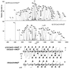
MS/MS spectra of ss12+ENU by (a) EPD of 3-• (one UV pulse, 5mJ; then 37% normalized collision energy, 30 ms), and (b) CID 3- (37% normalized collision energy, 30 ms). Precursor ions are noted with a star. Bolded and italicized product ions retain the modification. Not all of the EPD product ions are labeled due to the complexity of the spectrum. A summary of the product ions formed from EPD and CID for the ENU modified single strand 12-mer (c), in which the slash marks represent cleavages at that location. A triangle above the slash mark represents a product containing the ENU modification.
Scheme 1
Scheme 1
Oligodeoxynucleotide fragmentation nomenclature.
None
For modified oligodeoxynucleotides, UVPD at 193 nm followed by CID of the charge reduced species pinpoints the modification site more readily than CID alone.

Similar articles

Cited by

References

    1. Huber Christian, Oberacher Herbert. PCT Int. Appl. 2003:21.
    1. McLuckey ScottA, Van Berkel GaryJ, Glish GaryL. Journal of the American Society for Mass Spectrometry. 1992;3(1):60–70. - PubMed
    1. Keller KarinM, Brodbelt JenniferS. Analytical Biochemistry. 2004;326(2):200–210. - PubMed
    1. Guan Ziqiang, Kelleher NeilL, O'Connor PeterB, Aaserud DavidJ, Little DanielP, McLafferty FredW. International Journal of Mass Spectrometry and Ion Processes. 1996;157/158:357–364.
    1. Gabelica Valerie, Tabarin Thibault, Antoine Rodolphe, Rosu Frederic, Compagnon Isabelle, Broyer Michel, De Pauw Edwin, Dugourd Philippe. Analytical Chemistry. 2006;78(18):6564–6572. - PubMed

Publication types

MeSH terms

Substances