

Congenital ureteropelvic junction obstruction: human disease and animal models
- PMID: 20681980
- PMCID: PMC3101490
- DOI: 10.1111/j.1365-2613.2010.00727.x
Congenital ureteropelvic junction obstruction: human disease and animal models
Abstract
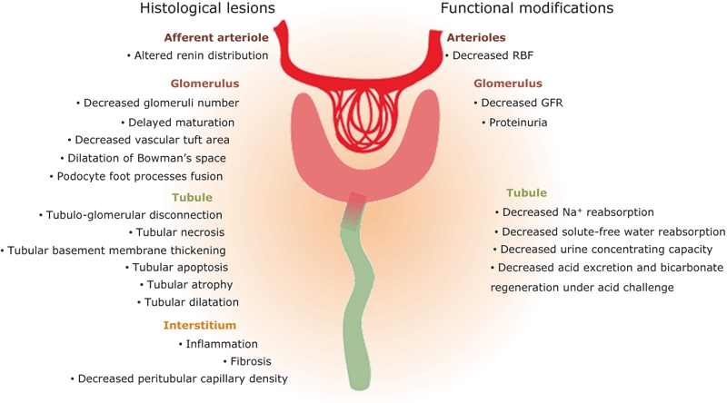
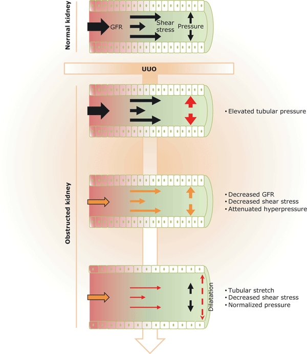
Ureteropelvic junction (UPJ) obstruction is the most frequently observed cause of obstructive nephropathy in children. Neonatal and foetal animal models have been developed that mimic closely what is observed in human disease. The purpose of this review is to discuss how obstructive nephropathy alters kidney histology and function and describe the molecular mechanisms involved in the progression of the lesions, including inflammation, proliferation/apoptosis, renin-angiotensin system activation and fibrosis, based on both human and animal data. Also we propose that during obstructive nephropathy, hydrodynamic modifications are early inducers of the tubular lesions, which are potentially at the origin of the pathology. Finally, an important observation in animal models is that relief of obstruction during kidney development has important effects on renal function later in adult life. A major short-coming is the absence of data on the impact of UPJ obstruction on long-term adult renal function to elucidate whether these animal data are also valid in humans.
© 2010 The Authors. Journal compilation © 2010 Blackwell Publishing Ltd.
Figures
Similar articles
-
Mechanisms of renal injury and progression of renal disease in congenital obstructive nephropathy.Pediatr Nephrol. 2010 Apr;25(4):687-97. doi: 10.1007/s00467-009-1316-5. Epub 2009 Oct 21. Pediatr Nephrol. 2010. PMID: 19844747 Review.
-
Variable chronic partial ureteral obstruction in the neonatal rat: a new model of ureteropelvic junction obstruction.Kidney Int. 2005 Jan;67(1):42-52. doi: 10.1111/j.1523-1755.2005.00052.x. Kidney Int. 2005. PMID: 15610226
-
Chronic partial ureteral obstruction and the developing kidney.Pediatr Radiol. 2008 Jan;38 Suppl 1:S35-40. doi: 10.1007/s00247-007-0585-z. Epub 2007 Dec 11. Pediatr Radiol. 2008. PMID: 18071697 Review.
-
Pathophysiology of obstructive nephropathy in the newborn.Semin Nephrol. 1998 Nov;18(6):585-93. Semin Nephrol. 1998. PMID: 9819149 Review.
-
iTRAQ-based proteomics and in vitro experiments reveals essential roles of ACE and AP-N in the renin-angiotensin system-mediated congenital ureteropelvic junction obstruction.Exp Cell Res. 2020 Aug 1;393(1):112086. doi: 10.1016/j.yexcr.2020.112086. Epub 2020 May 13. Exp Cell Res. 2020. PMID: 32416091
Cited by
-
Heterotaxy syndrome, dextrocardia, ureteropelvic obstruction, endometriosis, and pulmonary hypertension in an adult with congenital heart defects: a case report.J Med Case Rep. 2025 Jan 20;19(1):28. doi: 10.1186/s13256-025-05043-2. J Med Case Rep. 2025. PMID: 39833927 Free PMC article.
-
Developmental pathology of congenital kidney and urinary tract anomalies.Clin Kidney J. 2018 Dec 1;12(3):382-399. doi: 10.1093/ckj/sfy112. eCollection 2019 Jun. Clin Kidney J. 2018. PMID: 31198539 Free PMC article.
-
Proteomic urinary biomarker approach in renal disease: from discovery to implementation.Pediatr Nephrol. 2015 May;30(5):713-25. doi: 10.1007/s00467-014-2790-y. Epub 2014 Mar 15. Pediatr Nephrol. 2015. PMID: 24633400 Review.
-
Systems biology combining human- and animal-data miRNA and mRNA data identifies new targets in ureteropelvic junction obstruction.BMC Syst Biol. 2017 Mar 1;11(1):31. doi: 10.1186/s12918-017-0411-7. BMC Syst Biol. 2017. PMID: 28249581 Free PMC article.
-
The role of urinary TIMP1 and MMP9 levels in predicting vesicoureteral reflux in neonates with antenatal hydronephrosis.Pediatr Nephrol. 2014 May;29(5):871-8. doi: 10.1007/s00467-013-2693-3. Epub 2014 Jan 4. Pediatr Nephrol. 2014. PMID: 24389602
References
-
- Bajpai M, Bal CS, Tripathi M, Kalaivani M, Gupta AK. Prenatally diagnosed unilateral hydronephrosis: prognostic significance of plasma renin activity. J. Urol. 2007;178:2580–2584. - PubMed
-
- Basak A, Koch P, Dupelle M, et al. Inhibitory specificity and potency of proSAAS-derived peptides toward proprotein convertase 1. J. Biol. Chem. 2001;276:32720–32728. - PubMed
-
- Benfield MR, McDonald RA, Bartosh S, Ho PL, Harmon W. Changing trends in pediatric transplantation: 2001 Annual Report of the North American Pediatric Renal Transplant Cooperative Study. Pediatr. Transplant. 2003;7:321–335. - PubMed
-
- Benjannet S, Reudelhuber T, Mercure C, Rondeau N, Chretien M, Seidah NG. Proprotein conversion is determined by a multiplicity of factors including convertase processing, substrate specificity, and intracellular environment. Cell type-specific processing of human prorenin by the convertase PC1. J. Biol. Chem. 1992;267:11417–11423. - PubMed
Publication types
MeSH terms
LinkOut - more resources
Full Text Sources
Other Literature Sources
