Comparative Study
. 2010 Oct;91(5):460-71.
doi: 10.1111/j.1365-2613.2010.00725.x.

Distinct granuloma responses in C57BL/6J and BALB/cByJ mice in response to pristane

Affiliations
Comparative Study

Distinct granuloma responses in C57BL/6J and BALB/cByJ mice in response to pristane

Huaiyong Chen et al. Int J Exp Pathol. 2010 Oct.

Abstract

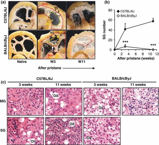
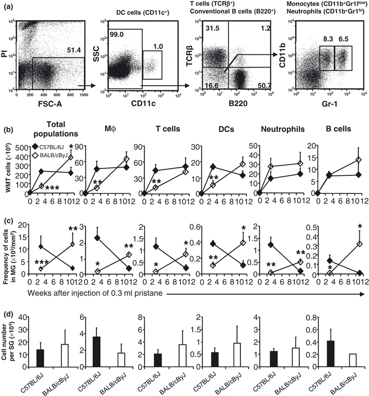
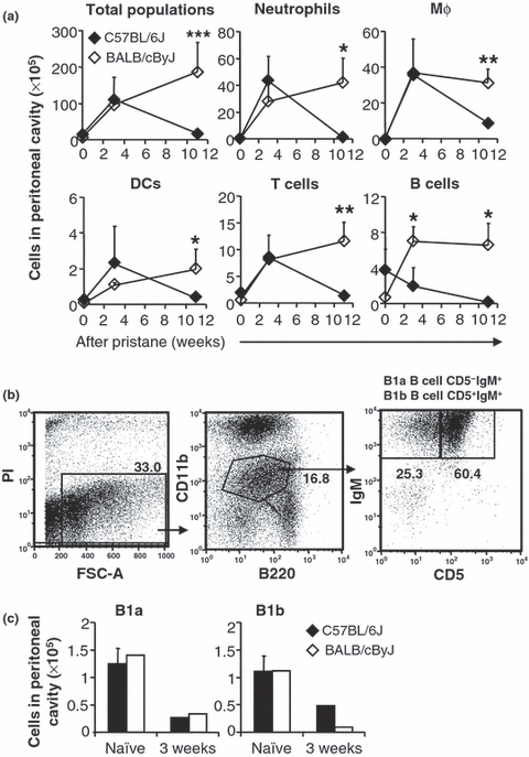
Granuloma formation is an inflammatory response of the host against invading pathogens or indigestible substances. We generated mesenteric oil granulomas by injecting pristane into the peritoneal cavity (PC) of mice, and compared oil granuloma formation in the C57BL/6J and BALB/cByJ strains of mice. The formation and kinetics of oil granulomas were distinct between the two strains. In C57BL/6J mice, injected pristane induced oil granuloma formation at both the mesenteric centers (MG) and margins (SG). MG was resolving by 11 weeks, and SG persisted. In BALB/cByJ mice, MG developed slower but persisted longer than in C57BL/6J mice, and SG resolved sooner than in C57BL/6J mice. Injection of India ink revealed that phagocytes were localised mainly to the SG in C57BL/6J mice, but were located diffusely in both MG and SG of BALB/cByJ mice. SG cells expressed more monocyte chemotactic protein-1 (MCP-1) mRNA than MG cells in C57BL/6J mice, but there was no difference in MCP-1 expression between the MG and SG in BALB/cByJ mice. These observations suggest that the recruitment of inflammatory leucocytes under the direction of chemokines differentiates the patterns of granuloma responses to pristane in C57BL/6J and BALB/cByJ mice.


Figures

Figure 1
Figure 1
Differential response of the mesentery to pristane in C57BL/6J and BALB/cByJ mice. (a) After i.p. injection of 300 μl (8.31 × 10⁻⁴ mol) of pristane, C57BL/6J and BALB/cByJ mice were sacrificed at week 3 or week 11, and gut associated WMT from naïve or pristane-primed mice was photographed. A white arrow indicates a polyp on mesentery. (b) SG were numerated in C57BL/6J and BALB/cByJ mice. ***P < 0.001 shows the difference between C57BL/6J mice vs. BALB/cByJ mice. (c) An equivalent area of MG or strip of SG was excised from either C57BL/6J or BALB/cByJ mice at week 3 or 11 after pristane, and the tissue was fixed and stained with hematoxylin and eosin solution. Representative pictures are shown. Original magnification (objective): ×63.
Figure 2
Figure 2
Leucocyte infiltration. (a) Identification of cell types by flow cytometry. Single cell suspensions prepared from spleen of pristane injected C57BL/6J mice (300 μl pristane, 11 weeks) were stained and analysed by flow cytometry as described in Materials and methods. Live cells were positively selected for analysis of CD11c expression, which results in identification of CD11c+ DCs and CD11c fraction. Analysing expression of B220 and TCRβ of CD11c cells reveals TCRβ+ fraction (T cells) and B220+ fraction (conventional B cells). The CD11cB220TCRβ fraction was furthered fractionated by expression of CD11b and Gr-1 into CD11b+Gr-1low (monocytes) and CD11b+Gr-1hi (neutrophils). Monocytes, after migrating into tissues, are called Mφ. Mφ in mesenteric tissue were identified as CD11b+Gr-1low. (b) Cellularity of WMT was analysed by flow cytometry. (c) MG was punched out with a 5-mm punch and its cellularity was determined as for the cellularity of WMT. Data represent the number of total cells of each cell type per 1 mm2 of MG. (d) 4–8 Individual SG were isolated with a 2-mm punch at week 3 after injection of pristane, and digested with collagenase. Cells were enumerated. The SG cellularity is presented as cell number/SG. Black diamonds or columns represent C57BL/6J mice, and white diamonds or columns represent BALB/cByJ mice. *P < 0.05; **P < 0.01; ***P < 0.001 shows the difference between C57BL/6J mice vs. BALB/cByJ mice.
Figure 3
Figure 3
Influx of inflammatory leucocytes into the PC following pristane injection. (a) Peritoneal cells were harvested from naïve or pristane injected C57BL/6J (black diamonds) and BALB/cByJ mice (white diamonds) at 3 weeks or 11 weeks. Numbers of total cells or leucocytes were isolated from peritoneal lavage, stained and analysed by flow cytometry. (b) In a separate study, peritoneal cells harvested at week 3 were labelled with B220-PE-Cy7, CD11b-APC-Cy7, CD5-PE and IgM-TxR to identify B1a cells (B220lowCD5+IgMhiCD11blow) and B1b cells (B220lowCD5IgMhiCD11blow). (c) The numbers of B1a and B1b in the PC are shown. Asterisks indicate significant differences between C57BL/6J and BALB/cByJ mice: *P < 0.05; **P < 0.01.
Figure 4
Figure 4
Characterisation of B cells in oil granulomas. (a) B cells in oil granulomas exhibit mature phenotypes (B220hiCD93-). WMT cells were recovered from naïve mice or mice injected with 300 μl pristane for 3 or 11 weeks, labeled with B220-PE-Cy7, CD93-APC and IgD-PE, and analysed by flow cytometry. BM cells from naïve C57BL/6J mice were harvested and labeled as indicative for mature conventional B cells (B220hiCD93IgD+) or immature B cells (B220lowCD93+IgD). (b) Plasma cells were only present in BALB/cByJ MG at week 11. A piece of MG, or a peripheral strip of mesentery containing SG, was excised from pristane-treated C57BL/6J and/or BALB/ cByJ mice (week 11), and embedded in OCT compound. Sections of 5-μm were cut, and fixed with acetone/methanol (1:1). Sections were stained with CD138-PE antibody, and visualised by fluorescent microscopy. Original magnification (objective): ×20. Arrows indicate positive staining.
Figure 5
Figure 5
India ink experiments. (a) C57BL/6J or BALB/cByJ mice were injected i.p. with 300 μl pristane, and 3 weeks later were injected i.p. with 0.5 ml of a 1/10 dilution of India ink in PBS. Twelve days after ink injection, the PC of mice was opened and gut-associated WMT was isolated and macro-photographed. Mice that were treated with only India ink were included as controls. (b) A representative picture of gut-associated WMT is shown. (c) Immunohistology of MG and SG. Sections were made from a block of peripheral mesentery containing SGs or an equivalent area of MG and were stained with biotin labelled CD11b, followed by incubation with streptavidin conjugated HRP. A piece of peripheral mesentery corresponding to SG (cSG) or a corresponding area of MG (cMG) in naïve mice was included as control. Original magnification (objective): ×20. (c) mRNA level of MCP-1 was determined in SG (Black columns) and MG (White columns) from C57BL/6J or BALB/cByJ mice. Asterisks indicate significant differences between SG and MG: *P < 0.05; **P < 0.01.

Similar articles

Cited by

References

    1. Bean AG, Roach DR, Briscoe H, et al. Structural deficiencies in granuloma formation in TNF gene-targeted mice underlie the heightened susceptibility to aerosol Mycobacterium tuberculosis infection, which is not compensated for by lymphotoxin. J. Immunol. 1999;162:3504–3511. - PubMed
    1. Birkness KA, Guarner J, Sable SB, et al. An in vitro model of the leukocyte interactions associated with granuloma formation in Mycobacterium tuberculosis infection. Immunol. Cell Biol. 2007;85:160–168. - PubMed
    1. Bowers RR, Houston F, Clinton R, Lewis M, Ballard R. A histological study of the carrageenan-induced granuloma in the rat lung. J. Pathol. 1980;132:243–253. - PubMed
    1. Chiu BC, Chensue SW. Chemokine responses in schistosomal antigen-elicited granuloma formation. Parasite Immunol. 2002;24:285–294. - PubMed
    1. Co DO, Hogan LH, Il-Kim S, Sandor M. T cell contributions to the different phases of granuloma formation. Immunol. Lett. 2004;92:135–142. - PubMed

Publication types

MeSH terms