. 2010 Aug 3:7:179.
doi: 10.1186/1743-422X-7-179.

Herpes simplex virus UL56 interacts with and regulates the Nedd4-family ubiquitin ligase Itch

Affiliations

Herpes simplex virus UL56 interacts with and regulates the Nedd4-family ubiquitin ligase Itch

Yoko Ushijima et al. Virol J. .

Abstract

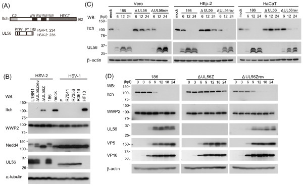
Background: Herpes simplex virus type 2 (HSV-2) is one of many viruses that exploits and modifies the cellular ubiquitin system. HSV-2 expresses the tegument protein UL56 that has been implicated in cytoplasmic transport and/or release of virions, and is a putative regulatory protein of Nedd4 ubiquitin ligase. In order to elucidate the biological function of UL56, this study examined the interaction of UL56 with the Nedd4-family ubiquitin ligase Itch and its role in the regulation of Itch. Additionally, we assessed the similarity between UL56 and regulatory proteins of Itch and Nedd4, Nedd4-family-interactins proteins (Ndfip).

Results: UL56 interacted with Itch, independent of additional viral proteins, and mediated more striking degradation of Itch, compared to Nedd4. Moreover, it was suggested that the lysosome pathway as well as the proteasome pathway was involved in the degradation of Itch. Other HSV-2 proteins with PY motifs, such as VP5 and VP16, did not mediate the degradation of endogenous Itch. Ndfip1 and Ndfip2 were similar in subcellular distribution patterns to UL56 and colocalized with UL56 in co-transfected cells.

Conclusions: We believe that this is the first report demonstrating the interaction of a HSV-specific protein and Itch. Thus, UL56 could function as a regulatory protein of Itch. The mechanism, function and significance of regulating Itch in HSV-2 infection remain unclear and warrant further investigation.


Figures

Figure 1
Figure 1
HSV infection causes a marked decrease of Itch in the presence of UL56. (A) Schematic representation of Itch and UL56. Itch (862 aa) contains a Ca2+/lipid binding C2 domain, four WW domains that interact with PY motifs, and a catalytic HECT domain. HSV UL56 (HSV-1, 234 aa; HSV-2, 235 aa) contains three PY motifs and a predicted transmembrane domain (TMD). (B) Infection with wild-type (HSV-1; F, HSV-2; 186) and various mutant HSV (HSV-1; US3-deletion mutant R7041, UL13 deletion mutant R7356, γ134.5-deletion mutant R3616, HSV-2; US3-deletion mutant L1BR1, UL56-reverted virus ΔUL56Zrev), but not infection with UL56-deficient HSV (HSV-1; HF10, HSV-2; ΔUL56Z), caused a marked decrease of Itch. Vero cells were mock-infected or infected with wild-type or mutant viruses and harvested at 24 hpi. Nedd4 changed only in cells infected with HSV-2 viruses except ΔUL56Z. WWP2, another Nedd4-family ubiquitin ligase, showed no remarkable change. (C-D) Itch decreases as HSV-2 infection proceeds in Vero, HEp-2 (C), and HaCaT (C, D) cells. Wild-type (186) and ΔUL56Zrev infection caused a marked decrease of Itch. In ΔUL56Z-infected cells, Itch was maintained at almost constant levels for up to 12 hpi. (D) VP5 and VP16 were detected similarly in cells infected with all three viruses. α-tubulin or β-actin were used as loading controls.
Figure 2
Figure 2
Effects of UL56 and other viral proteins with a PY motif on Itch. (A) Itch is markedly decreased in cells stably expressing UL56 (Vero-GFP-UL56). Lysates from Vero, Vero-GFP-UL56, or Vero-GFP cells were analyzed for Itch and other Nedd4-family ubiquitin ligases. Itch was markedly decreased in Vero-GFP-UL56 cells. (B) VP5 did not decrease endogenous Itch. Vero cells were transfected with plasmids encoding VP5 (pcDNA-VP5) or control plasmids (pcDNA3.1(+)). The levels of Itch did not change in cells transfected with pcDNA-VP5. (C) VP5 and VP16, but not ICP0, caused the decrease of exogenous Itch. Vero cells were co-transfected with plasmids encoding Itch (pcDNA-Itch) and plasmids encoding a viral protein (pMyc-ICP0, pcDNA-VP5, or pcDNA-VP16) or control plasmids (pCMV-Myc or pcDNA3.1(+)). The levels of Itch decreased in cells transfected with pcDNA-VP5 or pcDNA-VP16. (D) Overexpression of Itch has no effect on the protein levels of VP5, VP16, or ICP0. Vero cells were co-transfected with plasmids encoding a viral protein (pMyc-ICP0, pcDNA-VP5 or pcDNA-VP16) and either pcDNA-Itch or control plasmids (pcDNA-3.1(+)). The levels of viral proteins did not change with the overexpression of Itch. β-actin was used as a loading control.
Figure 3
Figure 3
Lysosome inhibitor and proteasome inhibitor block the decrease of Itch. (A) Itch expression is stable in the absence of UL56. Vero cells were cultured for 24 h with or without cycloheximide (CHX, 100 μg/ml) and the lysates were analyzed for Itch expression. Cyclin D3 was used as a control. (B) Vero cells were mock-treated or treated for 12 h with either MG132 (10 μM) or chloroquine (CQ, 100 μM) and infected with wild-type HSV-2. Itch was detected in both cells treated with MG132 and those with CQ at 12 hpi, but only in those with CQ at 24 hpi. (C) The decrease of Itch is blocked by a lysosome inhibitor and partially by a proteasome inhibitor in cells stably expressing UL56. Vero, Vero-GFP-UL56, or Vero-GFP cells were mock-treated or treated with either MG132 (10 μM) or CQ (100 μM) for 24 h. β-actin was used as a loading control.
Figure 4
Figure 4
UL56 interacts with Itch and changes the subcellular localization of Itch. Co-immunoprecipitation assay on HSV-2-infected cells (A) or stably UL56-expressing cells (B). (A)Vero cells were treated with chloroquine (CQ) for 12 h and subsequently mock-infected or infected with wild-type HSV-2 (186). Whole cell lysates (WCL) were immunoprecipitated (IP) with an anti-Itch antibody at 9 hpi. (B) WCL from Vero-GFP or Vero-GFP-UL56 cells treated with CQ for 24 h and immunoprecipitated with an anti-Itch antibody. UL56 was detected in the Itch-immunoprecipitates in HSV-2-infected cells (A) and stably UL56-expressing cells (B). β-actin was used as a loading control. Confocal immunofluorescence analysis of the subcellular localizations of Myc-Itch and UL56 in HSV-2-infected cells (C) or co-expressing cells (D). (C) HEp-2 cells were transfected with plasmids encoding Myc-Itch and subsequently infected with wild-type HSV-2. The Myc-Itch (red) showed the altered subcellular distribution with the reduced signal intensity after 6 hpi, when UL56 (green) became detectable. Myc-Itch colocalized with UL56 in the vesicular pattern. (D) HEp-2 cells were transfected with plasmids encoding UL56 (pcDNA-UL56) and/or pCMV-Myc-Itch. In co-expressing cells (bottom panels), Myc-Itch changed its distribution and colocalized with UL56 in the vesicular pattern. The Myc signal was reduced in co-expressing cells. Scale bars, 10 μm.
Figure 5
Figure 5
siRNA knockdown of Itch has no apparent effect on the viral growth. Vero cells were mock-transfected (RNA [-]) or transfected with either negative control siRNAs (siCont) or siRNAs specific to Itch (siItch) for 48 h (A), and subsequently infected with indicated viruses (B, C). (A) siItch efficiently and specifically down-regulates protein levels of Itch, whereas levels of Nedd4 and β-actin were constant. (B) A multi-step growth curve in siRNA-treated cells (MOI 0.003 PFU/ml). Both wild-type (186) and ΔUL56Z viruses showed similar growth kinetics in siCont-treated cells and siItch-treated cells. (C) Expression of Itch and viral proteins with PY motifs (VP5, VP16, and UL56) by immunoblot. There was no difference in viral protein level between siCont- and siItch-treated cells. β-actin was used as a loading control.
Figure 6
Figure 6
UL56 co-localizes with Ndfip1 and Ndfip2. (A) Schematic representation of UL56 and Ndfip proteins. HSV-2 UL56 (235 aa) contains three PY motifs and one predicted transmembrane domain (TMD). Ndfip1 (221 aa) and Ndfip2 (336 aa) contain two and three PY motifs respectively, and three TMD. (B-C) HEp-2 cells were transfected with plasmids encoding either Ndfip1-EGFP (pNdfip1-EGFP) (B) or Ndfip2-EGFP (pNdfip2-EGFP) (C) alone (left panels), or in combination with plasmids encoding UL56 (right panels). UL56 colocalized with Ndfip1-EGFP and Ndfip2-EGFP in co-expressing cells. (D-E) HEp-2 cells were transfected with pNdfip1-EGFP (D) or pNdfip2-EGFP (E), and subsequently infected with wild-type HSV-2. UL56 partially colocalized with Ndfip1-EGFP and Ndfip2-EGFP in infected cells. Scale bars, 10 μm.

Similar articles

Cited by

References

    1. Ball L. In: Fields Virology. 5. Knipe DM, Howley PM, editor. Vol. 1. Philadelphia: Lippincott Williams & Wilkin; 2007. Virus Replication Strategies; pp. 119–139.
    1. Isaacson MK, Ploegh HL. Ubiquitination, ubiquitin-like modifiers, and deubiquitination in viral infection. Cell Host Microbe. 2009;5:559–570. doi: 10.1016/j.chom.2009.05.012. - DOI - PMC - PubMed
    1. Randow F, Lehner PJ. Viral avoidance and exploitation of the ubiquitin system. Nat Cell Biol. 2009;11:527–534. doi: 10.1038/ncb0509-527. - DOI - PubMed
    1. Weissman AM. Themes and variations on ubiquitylation. Nat Rev Mol Cell Biol. 2001;2:169–178. doi: 10.1038/35056563. - DOI - PubMed
    1. Pickart CM. Mechanisms underlying ubiquitination. Annu Rev Biochem. 2001;70:503–533. doi: 10.1146/annurev.biochem.70.1.503. - DOI - PubMed

Publication types