

Crystallizing transmembrane peptides in lipidic mesophases
- PMID: 20682243
- PMCID: PMC2913208
- DOI: 10.1016/j.bpj.2010.05.011
Crystallizing transmembrane peptides in lipidic mesophases
Abstract
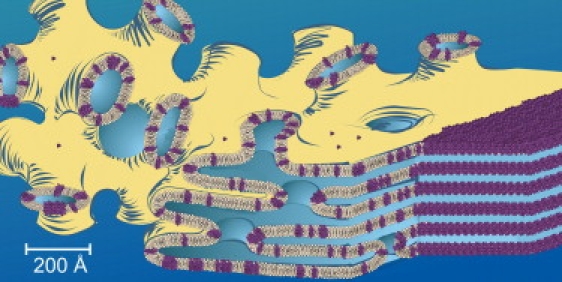
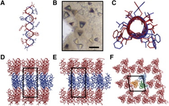
Structure determination of membrane proteins by crystallographic means has been facilitated by crystallization in lipidic mesophases. It has been suggested, however, that this so-called in meso method, as originally implemented, would not apply to small protein targets having </=4 transmembrane crossings. In our study, the hypothesis that the inherent flexibility of the mesophase would enable crystallogenesis of small proteins was tested using a transmembrane pentadecapeptide, linear gramicidin, which produced structure-grade crystals. This result suggests that the in meso method should be considered as a viable means for high-resolution structure determination of integral membrane peptides, many of which are predicted to be coded for in the human genome.
2010 Biophysical Society. Published by Elsevier Inc. All rights reserved.
Figures
Comment in
-
Modeling the membrane environment for membrane proteins.Biophys J. 2011 Apr 20;100(8):2073-4; author reply 2075. doi: 10.1016/j.bpj.2011.02.058. Biophys J. 2011. PMID: 21504744 Free PMC article. No abstract available.
Similar articles
-
Crystallizing membrane proteins using lipidic mesophases.Nat Protoc. 2009;4(5):706-31. doi: 10.1038/nprot.2009.31. Nat Protoc. 2009. PMID: 19390528 Free PMC article.
-
Crystallizing membrane proteins for structure determination: use of lipidic mesophases.Annu Rev Biophys. 2009;38:29-51. doi: 10.1146/annurev.biophys.050708.133655. Annu Rev Biophys. 2009. PMID: 19086821 Review.
-
Harvesting and cryo-cooling crystals of membrane proteins grown in lipidic mesophases for structure determination by macromolecular crystallography.J Vis Exp. 2012 Sep 2;(67):e4001. doi: 10.3791/4001. J Vis Exp. 2012. PMID: 22971942 Free PMC article.
-
Room to move: crystallizing membrane proteins in swollen lipidic mesophases.J Mol Biol. 2006 Apr 14;357(5):1605-18. doi: 10.1016/j.jmb.2006.01.049. Epub 2006 Feb 2. J Mol Biol. 2006. PMID: 16490208
-
A comprehensive review of the lipid cubic phase or in meso method for crystallizing membrane and soluble proteins and complexes.Acta Crystallogr F Struct Biol Commun. 2015 Jan 1;71(Pt 1):3-18. doi: 10.1107/S2053230X14026843. Epub 2015 Jan 1. Acta Crystallogr F Struct Biol Commun. 2015. PMID: 25615961 Free PMC article. Review.
Cited by
-
Crystallizing Membrane Proteins in Lipidic Mesophases. A Host Lipid Screen.Cryst Growth Des. 2011;11(2):530-537. doi: 10.1021/cg101378s. Cryst Growth Des. 2011. PMID: 21743796 Free PMC article.
-
Transmembrane Complexes of DAP12 Crystallized in Lipid Membranes Provide Insights into Control of Oligomerization in Immunoreceptor Assembly.Cell Rep. 2015 May 26;11(8):1184-92. doi: 10.1016/j.celrep.2015.04.045. Epub 2015 May 14. Cell Rep. 2015. PMID: 25981043 Free PMC article.
-
Crystallizing membrane proteins for structure-function studies using lipidic mesophases.Biochem Soc Trans. 2011 Jun;39(3):725-32. doi: 10.1042/BST0390725. Biochem Soc Trans. 2011. PMID: 21599641 Free PMC article.
-
Lipidic cubic phase technologies for membrane protein structural studies.Curr Opin Struct Biol. 2011 Aug;21(4):559-66. doi: 10.1016/j.sbi.2011.06.007. Epub 2011 Jul 19. Curr Opin Struct Biol. 2011. PMID: 21775127 Free PMC article. Review.
-
Methods of Measuring Mitochondrial Potassium Channels: A Critical Assessment.Int J Mol Sci. 2022 Jan 21;23(3):1210. doi: 10.3390/ijms23031210. Int J Mol Sci. 2022. PMID: 35163132 Free PMC article. Review.
References
MeSH terms
Substances
Associated data
- Actions
Grants and funding
LinkOut - more resources
Full Text Sources
