. 2010 Aug 4;99(3):790-7.
doi: 10.1016/j.bpj.2010.05.012.

Atomic force microscopy reveals the alternating subunit arrangement of the TRPP2-TRPV4 heterotetramer

Affiliations

Atomic force microscopy reveals the alternating subunit arrangement of the TRPP2-TRPV4 heterotetramer

Andrew P Stewart et al. Biophys J. .

Abstract

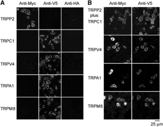
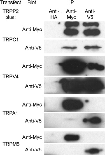
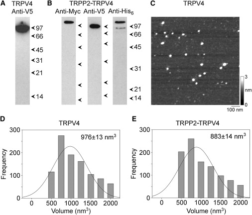
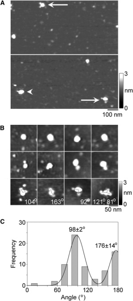
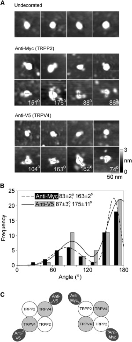
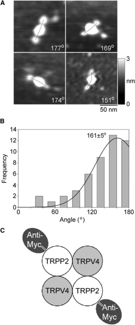
There is evidence that polycystin-2 (TRPP2) interacts with two other members of the transient receptor potential (TRP) family, TRPC1 and TRPV4. We have previously shown that TRPP2 forms a heteromeric complex with TRPC1, with a 2:2 stoichiometry and an alternating subunit arrangement. Here, we used coimmunoprecipitation to show that TRPP2 also interacts with TRPV4, but not with TRPA1 or TRPM8; hence, its promiscuity is limited. We then used atomic force microscopy to study the structure of the TRPV4 homomer and the interaction between TRPP2 and TRPV4. The molecular volume of V5-tagged TRPV4 isolated from singly-transfected tsA 201 cells indicated that it assembled as a homotetramer. The distribution of angles between pairs of anti-V5 antibodies bound to TRPV4 particles had a large peak close to 90 degrees and a smaller peak close to 180 degrees , again consistent with the assembly of TRPV4 as a homotetramer. In contrast, the angle distributions for decoration of the TRPP2-TRPV4 heteromer by either anti-Myc or anti-V5 antibodies had major peaks close to 180 degrees. This result indicates that TRPP2-TRPV4 assembles identically to TRPP2-TRPC1, suggesting a common subunit arrangement among heteromeric TRP channels.


Figures

Figure 1
Figure 1
Expression of TRP channels in tsA 201 cells. (A) Cells were transiently transfected with DNA encoding Myc/His6-tagged TRPP2 or V5/His6-tagged TRPV4, TRPC1, TRPA1, or TRPM8. Cells were fixed, permeabilized, and incubated with mouse monoclonal anti-Myc, anti-V5, or anti-HA (control) antibodies, followed by Cy3-conjugated goat anti-mouse secondary antibody. Cells were imaged by confocal laser scanning microscopy. (B) Cells were doubly transfected with TRPP2-Myc/His6 in addition to V5/His6-tagged TRPV4, TRPC1, TRPA1, or TRPM8. Cells were fixed, permeabilized, and incubated with rabbit polyclonal anti-Myc or mouse monoclonal anti-V5 antibodies, followed by Cy3-conjugated goat anti-rabbit or fluorescein isothiocyanate-conjugated goat anti-mouse secondary antibodies. As shown, the anti-Myc and anti-V5 signals extensively overlapped, indicating that the majority of transfected cells expressed both TRPP2 and its partner protein.
Figure 2
Figure 2
Coimmunoprecipitation of TRPP2 with other members of the TRP channel superfamily. TRPP2 was coexpressed with TRPC1, TRPV4, TRPA1, or TRPM8 by transient transfection of tsA 201 cells. Crude membrane fractions prepared from the cells were solubilized in 1% CHAPS. TRPP2 was immunoprecipitated using a rabbit polyclonal anti-Myc antibody, and its partner protein was immunoprecipitated using a mouse monoclonal anti-V5 antibody. A mouse monoclonal anti-HA antibody was used as a negative control. Immunoprecipitates were analyzed by SDS-polyacrylamide gel electrophoresis followed by immunoblotting using mouse monoclonal anti-Myc or anti-V5 antibodies. Immunoreactive bands were visualized using enhanced chemiluminescence.
Figure 3
Figure 3
Isolation and AFM imaging of TRPV4 homomers and TRPP2-TRPV4 heteromers. (A and B) Detection of proteins in samples from cells expressing either TRPV4 alone (A) or both TRPP2 and TRPV4 (B) eluted from Ni2+-agarose columns. Samples were analyzed by SDS-polyacrylamide gel electrophoresis and immunoblotting using anti-tag antibodies, followed by horseradish peroxidase-conjugated goat secondary antibodies. The total yield of isolated proteins was ∼500 ng, of which 30 ng was loaded on the gels. (Arrowheads) Molecular-mass markers (kDa). (C) Low-magnification AFM image of proteins isolated from TRPV4-expressing cells. (Right) Shade-height scale. (D and E) Frequency distributions of molecular volumes of proteins isolated from cells expressing TRPV4 (D) and TRPP2-TRPV4 (E). (Curves) Fitted Gaussian functions. The means of the distributions (± SE) are indicated.
Figure 4
Figure 4
Decoration of TRPV4 channels with anti-Myc antibodies. (A) Low-magnification AFM images of a sample of isolated TRPV4 that had been incubated with anti-Myc antibodies. (Arrowhead) Singly-decorated TRPV4 particle. (Arrows) Doubly-decorated particles. (Right) Shade-height scale. (B) Gallery of zoomed images of TRPV4 particles that are undecorated (top), or decorated by one (middle) or two (bottom) peripheral particles (antibodies). One triply decorated particle is also shown. Angles between pairs of antibodies are indicated. (Right) Shade-height scale. (C) Frequency distribution of angles between pairs of bound antibodies. The curve indicates the fitted Gaussian functions. The peaks of the distribution are indicated.
Figure 5
Figure 5
Decoration of TRPP2-TRPV4 channels with anti-Myc and anti-V5 antibodies. (A) Gallery of zoomed images of particles that are undecorated (top), decorated by one or two anti-Myc antibodies (middle), or decorated by one or two anti-V5 antibodies (bottom). Angles between pairs of antibodies are indicated. (Right) Shade-height scale. (B) Frequency distributions of angles between pairs of bound anti-Myc and anti-V5 antibodies. The curves indicate the fitted Gaussian functions. The peaks of the distributions are indicated. (C) (Diagram) Predominant subunit arrangement within the TRPP2-TRPV4 heteromer, as revealed by the antibody decoration patterns.
Figure 6
Figure 6
Decoration of TRPP2-TRPV4 channels isolated from cells expressing TRPP2-Myc and TRPV4-V5/His6 with anti-Myc antibodies. (A) Gallery of zoomed images of particles decorated by two anti-Myc antibodies. Angles between pairs of antibodies are indicated. (Right) Shade-height scale. (B) Frequency distributions of angles between pairs of bound anti-Myc antibodies. (Curve) Fitted Gaussian function. The peak of the distribution is indicated. (C) (Diagram) Subunit arrangement within the TRPP2-TRPV4 heteromer, as revealed by the antibody decoration pattern.

Similar articles

Cited by

References

    1. Mochizuki T., Wu G., Hayashi T., Somlo S. PKD2, a gene for polycystic kidney disease that encodes an integral membrane protein. Science. 1996;272:1339–1342. - PubMed
    1. Geng L., Boehmerle W., Somlo S. Syntaxin 5 regulates the endoplasmic reticulum channel-release properties of polycystin-2. Proc. Natl. Acad. Sci. USA. 2008;105:15920–15925. - PMC - PubMed
    1. Wegierski T., Steffl D., Köttgen M. TRPP2 channels regulate apoptosis through the Ca2+ concentration in the endoplasmic reticulum. EMBO J. 2009;28:490–499. - PMC - PubMed
    1. Hanaoka K., Qian F., Germino G.G. Co-assembly of polycystin-1 and -2 produces unique cation-permeable currents. Nature. 2000;408:990–994. - PubMed
    1. Nauli S.M., Alenghat F.J., Zhou J. Polycystins 1 and 2 mediate mechanosensation in the primary cilium of kidney cells. Nat. Genet. 2003;33:129–137. - PubMed

Publication types