

Role of cytoskeleton in controlling the disorder strength of cellular nanoscale architecture
- PMID: 20682278
- PMCID: PMC2913198
- DOI: 10.1016/j.bpj.2010.05.023
Role of cytoskeleton in controlling the disorder strength of cellular nanoscale architecture
Abstract
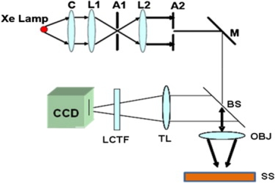
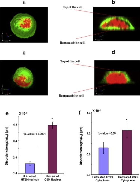
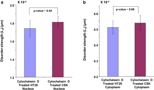
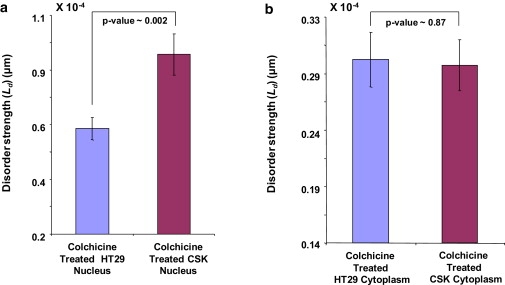
Cytoskeleton is ubiquitous throughout the cell and is involved in important cellular processes such as cellular transport, signal transduction, gene transcription, cell-division, etc. Partial wave spectroscopic microscopy is a novel optical technique that measures the statistical properties of cell nanoscale organization in terms of the disorder strength. It has been found previously that the increase in the disorder strength of cell nanoarchitecture is one of the earliest events in carcinogenesis. In this study, we investigate the cellular components responsible for the differential disorder strength between two morphologically (and hence microscopically) similar but genetically altered human colon cancer cell lines, HT29 cells and Csk shRNA-transfected HT29 cells that exhibit different degrees of neoplastic aggressiveness. To understand the role of cytoskeleton in nanoarchitectural alterations, we performed selective drug treatment on the specific cytoskeletal components of these cell types and studied the effects of cytoskeletal organization on disorder strength differences. We report that altering the cell nanoarchitecture by disrupting cytoskeletal organization leads to the attenuation of the disorder strength differences between microscopically indistinguishable HT29 and CSK constructs. We therefore demonstrate that cytoskeleton plays a role in the control of cellular nanoscale disorder.
2010 Biophysical Society. Published by Elsevier Inc. All rights reserved.
Figures
Similar articles
-
The role of the cytoskeleton in differentially regulating pressure-mediated effects on malignant colonocyte focal adhesion signaling and cell adhesion.Carcinogenesis. 2005 Oct;26(10):1687-97. doi: 10.1093/carcin/bgi135. Epub 2005 May 25. Carcinogenesis. 2005. PMID: 15917311
-
MEK/ERK pathway mediates cell-shape-dependent plasminogen activator inhibitor type 1 gene expression upon drug-induced disruption of the microfilament and microtubule networks.J Cell Sci. 2002 Aug 1;115(Pt 15):3093-103. doi: 10.1242/jcs.115.15.3093. J Cell Sci. 2002. PMID: 12118065
-
Role of the cytoskeleton in adhesion stabilization of human colorectal carcinoma cells to extracellular matrix components under dynamic conditions of laminar flow.Clin Exp Metastasis. 1999;17(8):713-21. doi: 10.1023/a:1006754829564. Clin Exp Metastasis. 1999. PMID: 10919716
-
Cytoskeleton-associated proteins: their role as cellular integrators in the neoplastic process.Crit Rev Oncol Hematol. 1985;3(3):191-204. doi: 10.1016/s1040-8428(85)80026-3. Crit Rev Oncol Hematol. 1985. PMID: 2412718 Review.
-
Role of c-Src in cellular events associated with colony-stimulating factor-1-induced spreading in osteoclasts.Mol Reprod Dev. 1997 Jan;46(1):104-8. doi: 10.1002/(SICI)1098-2795(199701)46:1<104::AID-MRD16>3.0.CO;2-2. Mol Reprod Dev. 1997. PMID: 8981371 Review.
Cited by
-
A Chemomechanical Model for Regulation of Contractility in the Embryonic Brain Tube.J Elast. 2021 Aug;145(1-2):77-98. doi: 10.1007/s10659-020-09811-7. Epub 2021 Jan 20. J Elast. 2021. PMID: 35400797 Free PMC article.
-
Macrogenomic engineering via modulation of the scaling of chromatin packing density.Nat Biomed Eng. 2017 Nov;1(11):902-913. doi: 10.1038/s41551-017-0153-2. Epub 2017 Nov 6. Nat Biomed Eng. 2017. PMID: 29450107 Free PMC article.
-
A computational framework for identifying cytoskeletal genes associated with age-related diseases.Sci Rep. 2025 Apr 26;15(1):14590. doi: 10.1038/s41598-025-97363-y. Sci Rep. 2025. PMID: 40287491 Free PMC article.
-
Light-scattering technologies for field carcinogenesis detection: a modality for endoscopic prescreening.Gastroenterology. 2011 Jan;140(1):35-41. doi: 10.1053/j.gastro.2010.11.023. Epub 2010 Nov 12. Gastroenterology. 2011. PMID: 21078318 Free PMC article.
-
Nanoscale markers of esophageal field carcinogenesis: potential implications for esophageal cancer screening.Endoscopy. 2013 Dec;45(12):983-8. doi: 10.1055/s-0033-1344617. Epub 2013 Sep 9. Endoscopy. 2013. PMID: 24019132 Free PMC article.
References
-
- Rao J.Y., Bonner R.B., Hemstreet G.P., 3rd Quantitative changes in cytoskeletal and nuclear actins during cellular transformation. Int. J. Cancer. 1997;70:423–429. - PubMed
-
- Ushijima T. Epigenetic field for cancerization. J. Biochem. Mol. Biol. 2007;40:142–150. - PubMed
-
- Alberts D.S., Einspahr J.G., Bartels P.H. Karyometry of the colonic mucosa. Cancer Epidemiol. Biomarkers Prev. 2007;16:2704–2716. - PubMed
-
- Chen L.C., Hao C.Y., Lee N.M. Alteration of gene expression in normal-appearing colon mucosa of APC(min) mice and human cancer patients. Cancer Res. 2004;64:3694–3700. - PubMed
-
- Hao C.Y., Moore D.H., Lee N.M. Altered gene expression in normal colonic mucosa of individuals with polyps of the colon. Dis. Colon Rectum. 2005;48:2329–2335. - PubMed
Publication types
MeSH terms
Substances
Grants and funding
LinkOut - more resources
Full Text Sources
