

The isolation, pathogenesis, diagnosis, transmission, and control of avian bornavirus and proventricular dilatation disease
- PMID: 20682432
- PMCID: PMC7110554
- DOI: 10.1016/j.cvex.2010.05.014
The isolation, pathogenesis, diagnosis, transmission, and control of avian bornavirus and proventricular dilatation disease
Abstract
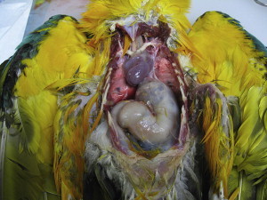
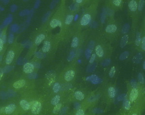
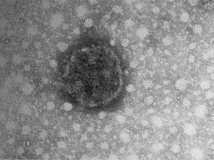
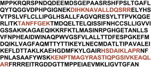
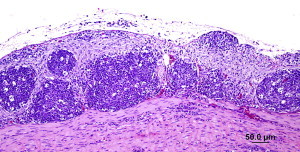
Proventricular dilatation disease (PDD) is a common infectious neurologic disease of birds comprising a dilatation of the proventriculus by ingested food as a result of defects in intestinal motility, which affects more than 50 species of psittacines, and is also known as Macaw wasting disease, neuropathic ganglioneuritis, or lymphoplasmacytic ganglioneuritis. Definitive diagnosis of PDD has been problematic due to the inconsistent distribution of lesions. Since its discovery, avian bornavirus (ABV) has been successfully cultured from the brains of psittacines diagnosed with PDD, providing a source of antigen for serologic assays and nucleic acid for molecular assays. This article provides evidence that ABV is the etiologic agent of PDD. Recent findings on the transmission, epidemiology, pathogenesis, diagnosis, and control of ABV infection and PDD are also reviewed.
Copyright 2010 Elsevier Inc. All rights reserved.
Figures
References
-
- Berhane Y., Binnington B., Hunter B. Peripheral neuritis in psittacine birds with proventricular dilation disease. Avian Pathol. 2001;73:563–570. - PubMed
-
- Gregory C., Ritchie B. Advances in understanding of proventricular dilation disease (PDD): detection of virus and viral nucleic acid in infected birds. Proc Annu Conf Assoc Avian Vet. 1999:41–43.
-
- Ritchie B. Epizootiology of proventricular dilatation disease in breeding cockatiels. Proc Annu Conf Assoc Avian Vet. 2004:41–45.
-
- Boutette J.B., Taylor M. Proventricular dilation disease: a review of research, literature, species differences, diagnostics, prognosis, and treatment. Proc Annu Conf Assoc Avian Vet. 2004:175–181.
-
- Shivaprasad H.L. Proventricular dilatation disease in a peregrine falcon. Proc Annu Conf Assoc Avian Vet. 2005:107–108.
Publication types
MeSH terms
LinkOut - more resources
Full Text Sources
