. 2010 Aug 6;7(2):163-73.
doi: 10.1016/j.stem.2010.05.019.

Adult SVZ lineage cells home to and leave the vascular niche via differential responses to SDF1/CXCR4 signaling

Affiliations

Adult SVZ lineage cells home to and leave the vascular niche via differential responses to SDF1/CXCR4 signaling

Erzsebet Kokovay et al. Cell Stem Cell. .

Abstract

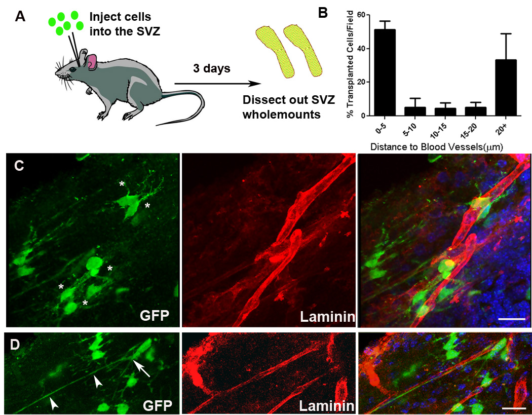
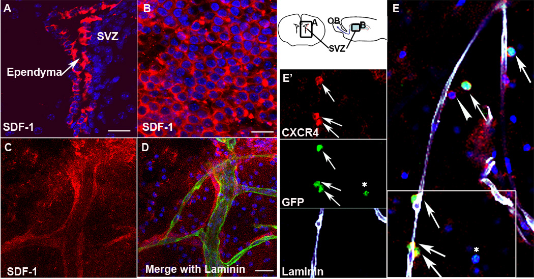
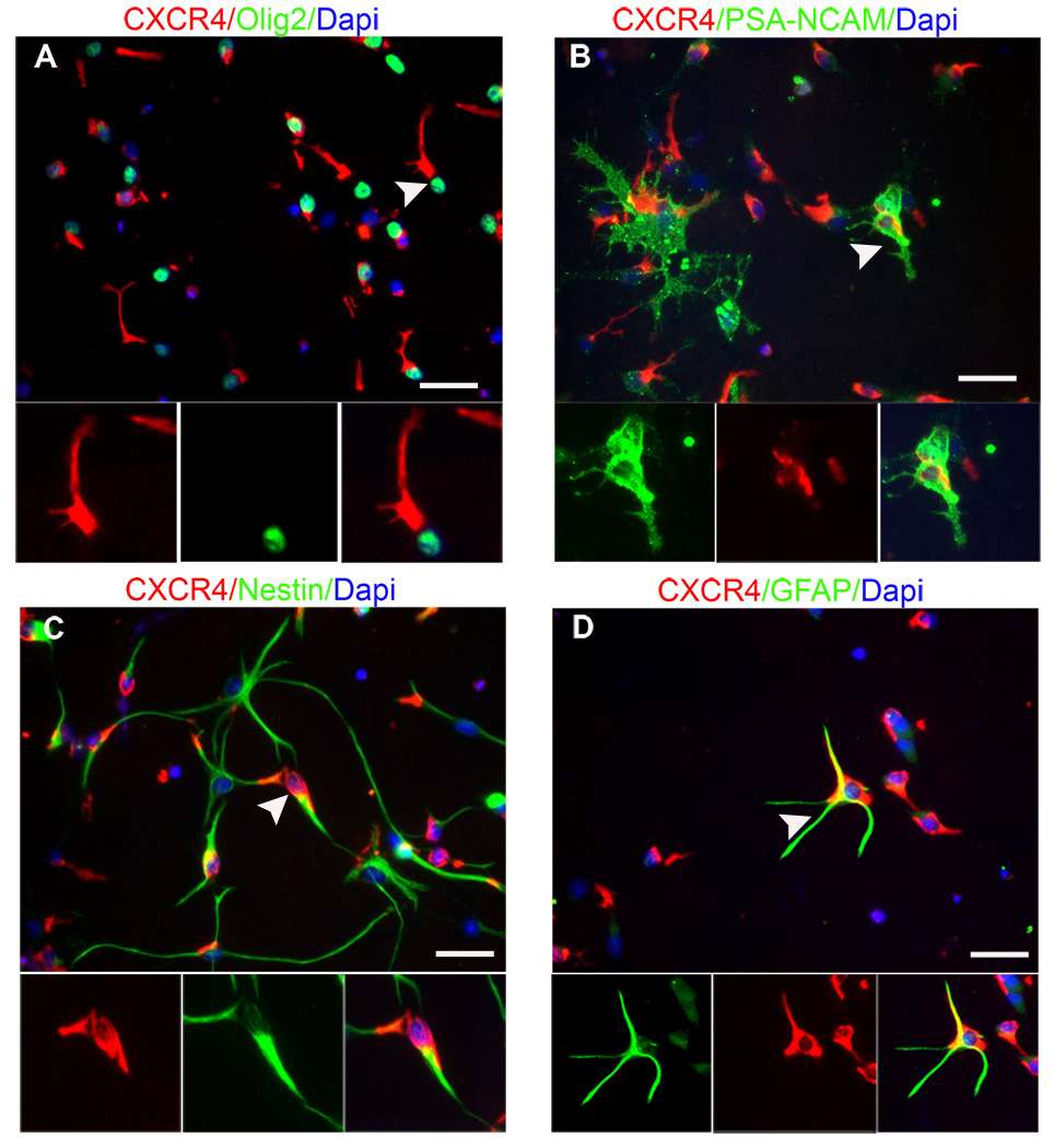
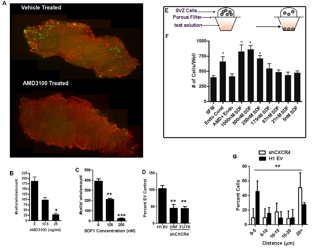
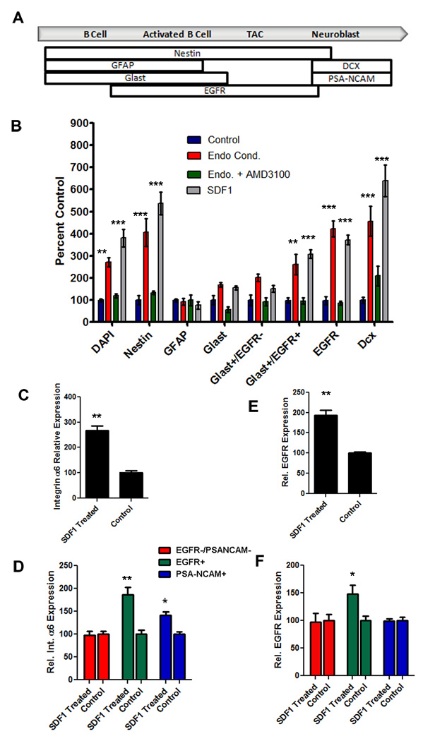
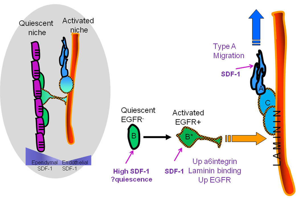
Neural progenitor cells (NPCs) in the adult subventricular zone (SVZ) are associated with ependymal and vasculature niches, which regulate stem cell self-renewal and differentiation. Activated Type B stem cells and their progeny, the transit-amplifying type C cells, which express EGFR, are most highly associated with vascular cells, indicating that this niche supports lineage progression. Here, we show that proliferative SVZ progenitor cells home to endothelial cells in a stromal-derived factor 1 (SDF1)- and CXC chemokine receptor 4 (CXCR4)-dependent manner. We show that SDF1 strongly upregulates EGFR and alpha6 integrin in activated type B and type C cells, enhancing their activated state and their ability to bind laminin in the vascular niche. SDF1 increases the motility of type A neuroblasts, which migrate from the SVZ toward the olfactory bulb. Thus, differential responses to SDF1 can regulate progenitor cell occupancy of and exit from the adult SVZ vascular niche.


Figures

Figure 1
Figure 1
NPCs associate with the SVZ vasculature following in vivo transplantation. (A) Schematic illustrating experimental design. NPCs were injected into the SVZ and then wholemounts were dissected for analysis by immunohistochemistry and confocal microscopy. (B) Quantification of the distance of transplanted cells to the nearest blood vessel surface. (C) Injected NPCs (green) are found near blood vessels (red); typical short processes of NPCs are indicated with asterisks. (D) NPCs with cell bodies lying directly on blood vessels (arrow) sometimes exhibit long processes extended along the blood vessel (arrowheads). Scale bars 20um.
Figure 2
Figure 2
NPCs preferentially associate with blood vessels in an organotypic wholemount assay system. (A) Schematic depicting the organotypic wholemount homing assay. The SVZ wholemount is microdissected from adult mice and cultured in membrane inserts. GFP+ cells are gently laid on the wholemount in a bubble of media and incubated, then processed for immunohistochemistry. Photomicrographs illustrating that NPCs (green) from a variety of sources enter the wholemounts and dispose along blood vessels (red): (B) endothelial cell expanded NPCs (C) Neurosphere-expanded NPCs. (D) Neurosphere expanded NPCs in the dorsal SVZ cluster into chains. Inset shows a higher magnification of (D) with nuclear Dapi stain in blue. (E) Acutely dissociated adult NPCs. (F) Quantification of the distance in microns of NPCs to blood vessels (the last bin represents 25 microns and greater). *p<0.001. Scale bars = 20um.
Figure 3
Figure 3
SDF1 and CXCR4 are expressed in the adult SVZ. SDF1 immunohistochemistry (red) shows strong expression in the ependymal cells of (A) a coronal section of the adult brain and (B) an organotypic wholemount. Schematic (right) indicates where pictures were taken. (C,D) SDF1 is expressed by blood vessels stained for laminin (green), and has a pattern consistent with a secreted gradient. (E) The SDF1 receptor CXCR4 (red) is expressed by transplanted NPCs (green) associated with the vasculature (white). Arrows indicate transplanted cells that express CXCR4. Arrowhead indicates an endogenous CXCR4 positive cell. Asterisk highlights a transplanted cell not associated with the vasculature that is CXCR4 negative. (E’) Individual channels from the boxed area in E. Scale bars = 20um
Figure 4
Figure 4
NPCs from the adult SVZ express CXCR4. Immunohistochemistry against the chemokine receptor CXCR4 in cultured NPCs after 1DIV reveals expression by a wide range of SVZ subtypes. (A) examples of olig2+ cells, a subpopulation of Type C cells, (B) PSA-NCAM, a Type A neuroblast marker (C) Nestin, a stem cell/progenitor marker and (D) GFAP, a stem/progenitor cell and astrocyte marker. Scale bars = 25um.
Figure 5
Figure 5
SDF1/CXCR4 signaling is important for NPCs to locate the SVZ vasculature. GFP+ NPCs were overlaid onto SVZ wholemounts, incubated then fixed. The vasculature was stained for laminin (red). The number of cells entering the wholemount was counted (A,B) AMD3100 treated cells show significantly reduced ability to integrate into the wholemount, and this was dose-responsive. (C) SDF1 added to the media overlying the wholemount disrupts the gradient and inhibits cell integration. (D) Quantification of the number of transplanted SVZ cells entering the wholemount following lentiviral knockdown of CXCR4 receptor. (E) Schematic of the chemotaxis chamber assay used to measure attraction of NPCs to various media. (F) The number of cells that migrated towards control serum free media (SFM), endothelial conditioned media (Endo Cond), endothelial conditioned media with 25ug/ml AMD3100 (Endo + AMD3100) or various concentration of the chemokine SDF1 in the media(*p <0.05; **p <0.001; ***p< 0.0001). (G) Quantification of the distance of transplanted cells transduced with shCXCR4 or H1 empty vector to nearest blood vessel surface **p<0.001 on the distribution of shCXCR4 cells versus EV cells.
Figure 6
Figure 6
Endothelial factors and SDF1 have differential effects on NPC populations. (A) Schematic showing the markers used to define the different populations of NPCs (modified from Pastrana et al., 2009). (B) Quantification of the types of NPCs that respond to endothelial conditioned media (red bars), endothelial conditioned media with AMD3100 (green bars) or 500nM SDF1 (grey bars) normalized to control (blue bars). (C) Expression levels of α6 integrin mRNA in acutely dissociated SVZ cells following treatment with 500nM SDF1 normalized to vehicle (D) a6 integrin expression levels in subpopulations of SVZ cells following SDF1 treatment. (E) EGFR expression in acutely dissociated SVZ cell population following treatment with SDF1 normalized to controls. (F) EGFR expression levels in subpopulations of SVZ cells following treatment with SDF1. *p<0.05, **p<0.001, ***p<0.0001
Figure 7
Figure 7
Summary model to describe the movement of SVZ NPCs to the vascular niche during lineage progression. SVZ blood vessels and ependymal cells (purple) represent two niches for SVZ stem cells - quiescent and activated, respectively. Both ependymal cells and endothelial cells secrete SDF1 creating a u-shaped gradient.CXCR4 is expressed on all stages of the SVZ lineage, but SDF1 has differential effects on the progenitor stages. High levels of SDF1 from ependymal cells stimulate quiescence of non-activated EGFR Type B cells (green). Upon activation and expression of EGFR (orange checked outline), SDF1 in the niche stimulates further upregulation of EGFR and α6 integrin that favor movement of the activated stem cells (green cell B*) towards the blood vessel surface, proliferation and generation of Type C cells (light blue cell). SDF1 also enhances migration of Type A cells (dark blue), which express lower levels of α6 integrin, thus promoting egress from the niche.

Comment in

References

    1. Aboody KS, Brown A, Rainov NG, Bower KA, Liu S, Yang W, Small JE, Herrlinger U, Ourednik V, Black PM, et al. Neural stem cells display extensive tropism for pathology in adult brain: evidence from intracranial gliomas. Proc Natl Acad Sci U S A. 2000;97:12846–12851. - PMC - PubMed
    1. Aguirre A, Rizvi TA, Ratner N, Gallo V. Overexpression of the epidermal growth factor receptor confers migratory properties to nonmigratory postnatal neural progenitors. J Neurosci. 2005;25:11092–11106. - PMC - PubMed
    1. Alvarez-Buylla A, Lim DA. For the long run: maintaining germinal niches in the adult brain. Neuron. 2004;41:683–686. - PubMed
    1. Arvidsson A, Collin T, Kirik D, Kokaia Z, Lindvall O. Neuronal replacement from endogenous precursors in the adult brain after stroke. Nat Med. 2002;8:963–970. - PubMed
    1. Ayuso-Sacido A, Moliterno JA, Kratovac S, Kapoor GS, O'Rourke DM, Holland EC, Garcia-Verdugo JM, Roy NS, Boockvar JA. Activated EGFR signaling increases proliferation, survival, and migration and blocks neuronal differentiation in post-natal neural stem cells. J Neurooncol. 2009 - PubMed

Publication types

MeSH terms