

Decoupling diffusional from dimensional control of signaling in 3D culture reveals a role for myosin in tubulogenesis
- PMID: 20682635
- PMCID: PMC2923566
- DOI: 10.1242/jcs.055079
Decoupling diffusional from dimensional control of signaling in 3D culture reveals a role for myosin in tubulogenesis
Abstract
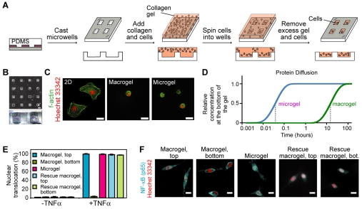
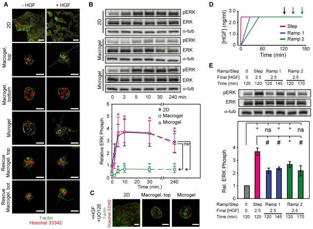
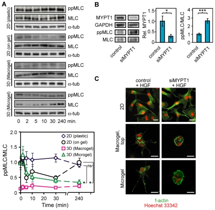
We present a novel microfabricated platform to culture cells within arrays of micrometer-scale three-dimensional (3D) extracellular matrix scaffolds (microgels). These microscale cultures eliminate diffusion barriers that are intrinsic to traditional 3D culture systems (macrogels) and enable uniform cytokine stimulation of the entire culture population, as well as allow immunolabeling, imaging and population-based biochemical assays across the relatively coplanar microgels. Examining early signaling associated with hepatocyte growth factor (HGF)-mediated scattering and tubulogenesis of MDCK cells revealed that 3D culture modulates cellular responses both through dimensionality and altered stimulation rates. Comparing responses in 2D culture, microgels and macrogels demonstrated that HGF-induced ERK signaling was driven by the dynamics of stimulation and not by whether cells were in a 2D or 3D environment, and that this ERK signaling was equally important for HGF-induced cell scattering on 2D substrates and tubulogenesis in 3D. By contrast, we discovered a specific HGF-induced increase in myosin expression leading to sustained downregulation of myosin activity that occurred only within 3D contexts and was required for 3D tubulogenesis but not 2D scattering. Interestingly, although absent in cells on collagen-coated plates, downregulation of myosin activity also occurred for cells on collagen gels, but was transient and mediated by a combination of myosin dephosphorylation and enhanced myosin expression. Furthermore, upregulating myosin activity via siRNA targeted to a myosin phosphatase did not attenuate scattering in 2D but did inhibit tubulogenesis in 3D. Together, these results demonstrate that cellular responses to soluble cues in 3D culture are regulated by both rates of stimulation and by matrix dimensionality, and highlight the importance of decoupling these effects to identify early signals relevant to cellular function in 3D environments.
Figures
Similar articles
-
Microscopic analysis of the cellular events during scatter factor/hepatocyte growth factor-induced epithelial tubulogenesis.J Anat. 2003 Nov;203(5):483-503. doi: 10.1046/j.1469-7580.2003.00238.x. J Anat. 2003. PMID: 14635802 Free PMC article.
-
Hepatocyte growth factor upregulates alpha2beta1 integrin in Madin-Darby canine kidney cells: implications in tubulogenesis.J Biomed Sci. 2002 May-Jun;9(3):261-72. doi: 10.1007/BF02256073. J Biomed Sci. 2002. PMID: 12065901
-
Novel MAPK-dependent and -independent tubulogenes identified via microarray analysis of 3D-cultured Madin-Darby canine kidney cells.Am J Physiol Renal Physiol. 2014 May 1;306(9):F1047-58. doi: 10.1152/ajprenal.00589.2013. Epub 2014 Feb 26. Am J Physiol Renal Physiol. 2014. PMID: 24573390 Free PMC article.
-
Bridging the Gap: From 2D Cell Culture to 3D Microengineered Extracellular Matrices.Adv Healthc Mater. 2015 Dec 30;4(18):2780-96. doi: 10.1002/adhm.201500427. Epub 2015 Nov 23. Adv Healthc Mater. 2015. PMID: 26592366 Free PMC article. Review.
-
Dimensions and dynamics in integrin function.Braz J Med Biol Res. 2003 Aug;36(8):959-66. doi: 10.1590/s0100-879x2003000800001. Epub 2003 Jul 23. Braz J Med Biol Res. 2003. PMID: 12886449 Review.
Cited by
-
A potential role for differential contractility in early brain development and evolution.Biomech Model Mechanobiol. 2012 Nov;11(8):1251-62. doi: 10.1007/s10237-012-0389-4. Epub 2012 Mar 31. Biomech Model Mechanobiol. 2012. PMID: 22466353 Free PMC article.
-
Regulation of Epithelial-to-Mesenchymal Transition Using Biomimetic Fibrous Scaffolds.ACS Appl Mater Interfaces. 2016 Jul 20;8(28):17915-26. doi: 10.1021/acsami.6b05646. Epub 2016 Jul 5. ACS Appl Mater Interfaces. 2016. PMID: 27322677 Free PMC article.
-
Novel insights from 3D models: the pivotal role of physical symmetry in epithelial organization.Sci Rep. 2015 Oct 16;5:15153. doi: 10.1038/srep15153. Sci Rep. 2015. PMID: 26472542 Free PMC article.
-
One-dimensional patterning of cells in silicone wells via compression-induced fracture.J Biomed Mater Res A. 2014 May;102(5):1361-9. doi: 10.1002/jbm.a.34814. Epub 2013 Jun 11. J Biomed Mater Res A. 2014. PMID: 23733484 Free PMC article.
-
Fracture-based micro- and nanofabrication for biological applications.Biomater Sci. 2014 Mar 1;2(3):288-296. doi: 10.1039/C3BM60276A. Biomater Sci. 2014. PMID: 24707353 Free PMC article.
References
-
- Birgersdotter A., Sandberg R., Ernberg I. (2005). Gene expression perturbation in vitro-a growing case for three-dimensional (3D) culture systems. Semin. Cancer Biol. 15, 405-412 - PubMed
-
- Bollenbach T., Pantazis P., Kicheva A., Bokel C., Gonzalez-Gaitan M., Julicher F. (2008). Precision of the Dpp gradient. Development 135, 1137-1146 - PubMed
-
- Chen C. S., Mrksich M., Huang S., Whitesides G. M., Ingber D. E. (1997). Geometric control of cell life and death. Science 276, 1425-1428 - PubMed
-
- Cukierman E., Pankov R., Yamada K. M. (2002). Cell interactions with three-dimensional matrices. Curr. Opin. Cell Biol. 14, 633-639 - PubMed
Publication types
MeSH terms
Substances
Grants and funding
LinkOut - more resources
Full Text Sources
Miscellaneous
