Basal body movements orchestrate membrane organelle division and cell morphogenesis in Trypanosoma brucei
- PMID: 20682637
- PMCID: PMC2923567
- DOI: 10.1242/jcs.074161
Basal body movements orchestrate membrane organelle division and cell morphogenesis in Trypanosoma brucei
Abstract
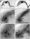
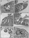
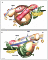
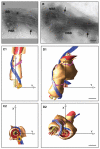
The defined shape and single-copy organelles of Trypanosoma brucei mean that it provides an excellent model in which to study how duplication and segregation of organelles is interfaced with morphogenesis of overall cell shape and form. The centriole or basal body of eukaryotic cells is often seen to be at the centre of such processes. We have used a combination of electron microscopy and electron tomography techniques to provide a detailed three-dimensional view of duplication of the basal body in trypanosomes. We show that the basal body duplication and maturation cycle exerts an influence on the intimately associated flagellar pocket membrane system that is the portal for secretion and uptake from this cell. At the start of the cell cycle, a probasal body is positioned anterior to the basal body of the existing flagellum. At the G1-S transition, the probasal body matures, elongates and invades the pre-existing flagellar pocket to form the new flagellar axoneme. The new basal body undergoes a spectacular anti-clockwise rotation around the old flagellum, while its short new axoneme is associated with the pre-existing flagellar pocket. This rotation and subsequent posterior movements results in division of the flagellar pocket and ultimately sets parameters for subsequent daughter cell morphogenesis.
Figures


Similar articles
-
Proximity Interactions among Basal Body Components in Trypanosoma brucei Identify Novel Regulators of Basal Body Biogenesis and Inheritance.mBio. 2017 Jan 3;8(1):e02120-16. doi: 10.1128/mBio.02120-16. mBio. 2017. PMID: 28049148 Free PMC article.
-
Basal body structure and cell cycle-dependent biogenesis in Trypanosoma brucei.Cilia. 2016 Feb 8;5:5. doi: 10.1186/s13630-016-0023-7. eCollection 2015. Cilia. 2016. PMID: 26862392 Free PMC article. Review.
-
The kinetoplast duplication cycle in Trypanosoma brucei is orchestrated by cytoskeleton-mediated cell morphogenesis.Mol Cell Biol. 2011 Mar;31(5):1012-21. doi: 10.1128/MCB.01176-10. Epub 2010 Dec 20. Mol Cell Biol. 2011. PMID: 21173163 Free PMC article.
-
Clathrin-dependent targeting of receptors to the flagellar pocket of procyclic-form Trypanosoma brucei.Eukaryot Cell. 2004 Aug;3(4):1004-14. doi: 10.1128/EC.3.4.1004-1014.2004. Eukaryot Cell. 2004. PMID: 15302833 Free PMC article.
-
The Flagellum Attachment Zone: 'The Cellular Ruler' of Trypanosome Morphology.Trends Parasitol. 2016 Apr;32(4):309-324. doi: 10.1016/j.pt.2015.12.010. Epub 2016 Jan 8. Trends Parasitol. 2016. PMID: 26776656 Free PMC article. Review.
Cited by
-
An analogue-sensitive approach identifies basal body rotation and flagellum attachment zone elongation as key functions of PLK in Trypanosoma brucei.Mol Biol Cell. 2013 May;24(9):1321-33. doi: 10.1091/mbc.E12-12-0846. Epub 2013 Feb 27. Mol Biol Cell. 2013. PMID: 23447704 Free PMC article.
-
A Trypanosoma brucei protein required for maintenance of the flagellum attachment zone and flagellar pocket ER domains.Protist. 2012 Jul;163(4):602-15. doi: 10.1016/j.protis.2011.10.010. Epub 2011 Dec 18. Protist. 2012. PMID: 22186015 Free PMC article.
-
TAC102 Is a Novel Component of the Mitochondrial Genome Segregation Machinery in Trypanosomes.PLoS Pathog. 2016 May 11;12(5):e1005586. doi: 10.1371/journal.ppat.1005586. eCollection 2016 May. PLoS Pathog. 2016. PMID: 27168148 Free PMC article.
-
Polo-like kinase in trypanosomes: an odd member out of the Polo family.Open Biol. 2020 Oct;10(10):200189. doi: 10.1098/rsob.200189. Epub 2020 Oct 14. Open Biol. 2020. PMID: 33050792 Free PMC article.
-
A specific basal body linker protein provides the connection function for basal body inheritance in trypanosomes.Proc Natl Acad Sci U S A. 2021 Feb 23;118(8):e2014040118. doi: 10.1073/pnas.2014040118. Proc Natl Acad Sci U S A. 2021. PMID: 33597294 Free PMC article.
References
-
- Absalon S., Blisnick T., Bonhivers M., Kohl L., Cayet N., Toutirais G., Buisson J., Robinson D., Bastin P. (2008). Flagellum elongation is required for correct structure, orientation and function of the flagellar pocket in Trypanosoma brucei. J. Cell Sci. 121, 3704-3716 - PubMed
Publication types
MeSH terms
Grants and funding
LinkOut - more resources
Full Text Sources

