. 2010 Nov;59(11):2799-808.
doi: 10.2337/db09-1478. Epub 2010 Aug 3.

INS-1 cells undergoing caspase-dependent apoptosis enhance the regenerative capacity of neighboring cells

Affiliations

INS-1 cells undergoing caspase-dependent apoptosis enhance the regenerative capacity of neighboring cells

Caroline Bonner et al. Diabetes. 2010 Nov.

Abstract

Objective: In diabetes, β-cell mass is not static but in a constant process of cell death and renewal. Inactivating mutations in transcription factor 1 (tcf-1)/hepatocyte nuclear factor1a (hnf1a) result in decreased β-cell mass and HNF1A-maturity onset diabetes of the young (HNF1A-MODY). Here, we investigated the effect of a dominant-negative HNF1A mutant (DN-HNF1A) induced apoptosis on the regenerative capacity of INS-1 cells.

Research design and methods: DN-HNF1A was expressed in INS-1 cells using a reverse tetracycline-dependent transactivator system. Gene(s)/protein(s) involved in β-cell regeneration were investigated by real-time quantitative RT-PCR, Western blotting, and immunohistochemistry. Pancreatic stone protein/regenerating protein (PSP/reg) serum levels in human subjects were detected by enzyme-linked immunosorbent assay.

Results: We detected a prominent induction of PSP/reg at the gene and protein level during DN-HNF1A-induced apoptosis. Elevated PSP/reg levels were also detected in islets of transgenic HNF1A-MODY mice and in the serum of HNF1A-MODY patients. The induction of PSP/reg was glucose dependent and mediated by caspase activation during apoptosis. Interestingly, the supernatant from DN-HNF1A-expressing cells, but not DN-HNF1A-expressing cells treated with zVAD.fmk, was sufficient to induce PSP/reg gene expression and increase cell proliferation in naïve, untreated INS-1 cells. Further experiments demonstrated that annexin-V-positive microparticles originating from apoptosing INS-1 cells mediated the induction of PSP/reg. Treatment with recombinant PSP/reg reversed the phenotype of DN-HNF1A-induced cells by stimulating cell proliferation and increasing insulin gene expression.

Conclusions: Our results suggest that apoptosing INS-1 cells shed microparticles that may stimulate PSP/reg induction in neighboring cells, a mechanism that may facilitate the recovery of β-cell mass in HNF1A-MODY.


Figures
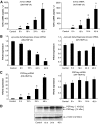
FIG. 1.
FIG. 1.
The inducible expression of DN-HNF1A leads to a potent induction of PSP/reg mRNA and protein. A: The time course of the hnf1a gene induction in DN-HNF1A and WT-HNF1A induced INS-1 cells, represented by absolute quantitative PCR (qPCR). Data are presented as cDNA copy number per microliter. Data are represented as means ± SE from n = 3 cultures. The experiment was repeated three times with similar results. *P < 0.05 indicates the difference from noninduced controls. B: Quantification of the pyruvate dehydrogenase kinase (Pdk1) gene expression. DN-HNF1A and WT-HNF1A INS-1 cells were induced with doxycycline from 0 to 48 h. Pdk1 mRNA expression was examined using real-time qPCR relative to β-actin. Data shown are the means ± SEM of n = 3 cultures. The experiment was repeated four times with similar results. *P < 0.05, difference from noninduced controls. C: Quantification of PSP/reg gene expression following DN-HNF1A and WT-HNF1A induction in INS-1 cells. INS-1 cells were treated with 500 ng/ml doxycycline from 0 to 48 h. mRNA expression of PSP/reg was examined using real-time qPCR relative to β-actin. Expression levels were normalized to control cells and data represent means ± SEM from n = 3 cultures. *P < 0.05, difference from noninduced controls. Experiments were repeated six times with similar results (D) whole cell lysates (25 μg) were analyzed by Western blotting on 15% SDS-PAGE. Membranes were probed with a polyclonal antibody recognizing PSP/reg. The 14-kDa fragment represents a PSP/reg cleavage product. β-Actin served as a loading control. Approximate molecular weights are provided on the right side of the figure. Similar results were obtained in two separate experiments.
FIG. 2.
FIG. 2.
High extracellular glucose potentiates PSP/reg mRNA induction in DN-HNF1A expressing INS-1 cells. A: INS-1 cells were induced to express DN-HNF1A for 24 h. Cultures were continued for 2 h at either 3, 6, 12, and 18 mmol/l glucose concentrations. After this period, mRNA expression of PSP/reg was analyzed by real-time qPCR. Expression levels were normalized to noninduced control cells and data are represented as means ± SEM from n = 3 cultures. *P < 0.05, difference from non induced controls; #P < 0.05, difference from induced cultures at 3 mmol/l glucose. Experiment was repeated three times with similar results. B: PSP/reg immunoreactivity in islet cells in an in vivo mouse model of DN-HNF1A–induced apoptosis. Paraffin-embedded pancreatic sections on slides from 5-month-old wild-type C57BL/6JBomTac control mice and RIP-DN-HNF1A transgenic mice were double stained using antibodies against PSP/reg and insulin. Primary antibodies were recognized by secondary antibodies coupled to Rhodamine (red) for PSP/reg and to FITC (green) for insulin, respectively. Cell nuclei were stained with DAPI (blue). Bar = 10 μm. C: PSP/reg immunoreactivity in islets cells in proximity to cells showing apoptotic nuclear morphology. High magnification images of PSP/reg immunoreactivity and DAPI staining in islets of DN-HNF1A mice. Arrows indicate cells exhibiting apoptotic nuclear morphology. Bar = 10 μm. D: PSP/reg protein is detectable in the serum of HNF1A-MODY subjects and type 1 diabetic subjects. Serum from HNF1A-MODY patients, their MODY-negative family members, and type 1 diabetic patients were analyzed by a specific ELISA detecting human PSP/reg. The square indicates the normal range (R.G., historic data). Individual values, the average as well as the SD are plotted. PSP/reg levels in HNF1A-MODY and type 1 diabetic subjects were significantly different from MODY-negative, control family members (*P < 0.05). (A high-quality digital representation of this figure is available in the online issue.)
FIG. 3.
FIG. 3.
Activation of caspases during DN-HNF1A–induced apoptosis triggers PSP/reg induction. A: Time course of caspase-3–like protease activity in whole-cell extracts. INS-1 cells were induced to overexpress WT-HNF1A or DN-HNF1A for 0, 16, 24, and 48 h. As a control, parental INS-1 cells were treated with doxycycline for 0, 16, 24, and 48 h. Caspase protease activity was measured by cleavage of the fluorigenic substrate Ac-DEVD-AMC (10 μmol/l). Activities are represented as increase in AMC fluorescence (in AU) per 1 h per microgram of protein. Data represent means ± SE from n = 6 cultures. Experiments were repeated two times with similar results. *P < 0.05, difference from noninduced controls. B: Treatment with the broad spectrum caspase inhibitor zVAD.fmk (100 μmol/l) inhibits apoptosis after induction of DN-HNF1A. Cultures were simultaneously pretreated with doxycycline and zVAD.fmk or vehicle (DMSO; zVAD.fmk) for 48. Apoptotic cell morphology was assessed by DAPI staining of nuclear chromatin. C: zVAD.fmk inhibits PSP/reg mRNA induction. The mRNA expression of PSP/reg was examined using real-time qPCR. Expression levels were normalized to noninduced plus noninduced zVAD.fmk–treated control cells. Data are represented as means ± SE from n = 3 cultures.*P < 0.05, difference from noninduced controls. #P < 0.05, difference compared with doxycycline alone–treated cultures. The experiment was repeated three times with similar results. D: PSP/reg protein expression was detected by Western blotting. A duplicate experiment showed similar results. Membrane was stripped and reprobed with anti–β-actin as a loading control. E: PSP/reg gene induction in parental INS-1 cells treated with etoposide is caspase dependent. INS-1 cells were simultaneously treated with etoposide (20 μmol/l) or vehicle (0.1% DMSO) for 24 h in the presence and absence of zVAD.fmk (100 μmol/l). Following treatment, mRNA expression of PSP/reg was examined using real-time qPCR. Expression levels were normalized to control cells. Data are represented as means ± SE from n = 3 separate cultures. *P < 0.05, difference from non induced controls. #P < 0.05, difference compared with etoposide alone–treated cultures. Data shown are the mean of n = 3 separate experiments.
FIG. 4.
FIG. 4.
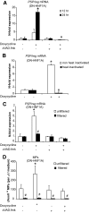
Conditioned medium from DN-HNF1A–induced INS-1 cells results in a prominent increase in PSP/reg mRNA expression in naïve INS1 Cells. A: INS-1 cells were induced with 500 ng/ml of doxycycline in the presence and absence of zVAD.fmk (100 μmol/l) at 0.05% serum in 6 mmol/l glucose for 48 h. The conditioned culture medium was added to the noninduced, naive cells for a further 24 h. After treatment, mRNA expression of PSP/reg was analyzed by real-time qPCR. Expression levels were normalized to noninduced controls. Data are represented as means ± SE from n = 3 cultures. *P < 0.05, difference from noninduced controls; #P < 0.05, difference from doxycycline alone–treated cultures. Experiments were repeated six times with similar results. B: Duplicate experiments of INS-1 cells were induced for 48 h. Conditioned culture medium was heat inactivated at 95°C for 15 min and added to the naive cells for a further 24 h. PSP/reg gene expression was analyzed by real-time qPCR. Data are represented as means ± SE from n = 3 cultures. *P < 0.05, difference from noninduced controls. Experiments were repeated four times with similar results. C: The conditioned medium from cells treated as described in A above was centrifuged and filtered through 0.20 μm filters and added to naïve INS-1 cells for 24 h. Unfiltered conditioned medium served as a control. PSP/reg gene expression was analyzed by real-time qPCR. Data are represented as means ± SE from n = 3 cultures. *P < 0.05, difference from noninduced controls. Experiments were repeated three times with similar results. D: INS-1 cells were induced with doxycycline in the presence and absence of zVAD.fmk. The conditioned culture medium was added to naive cells for a further 24 h. Supernatants were collected and detached cells were precipitated at (300g), followed by additional 800g centrifugation. Microparticles were quantified as described in research design and methods.
FIG. 5.
FIG. 5.
Treatment of INS-1 cells with recombinant PSP/reg protein reverses the phenotype of HNF1A-MODY cells. A: INS-1 cells were induced to express DN-HNF1A for 24 h before being cultured for a further 24 h in the presence and absence of 10 ng/ml of rPSP/reg for 24 h. Proliferation was assessed by BrdU incorporation into the cells. Data are a means ± SE from n = 6 cultures. Experiments were repeated three times with similar results. *P < 0.05, difference from noninduced controls. #P < 0.05, difference compared with doxycycline alone–treated cultures. B and C: DN-HNF1A INS-1 cells were treated as described in A above. Insulin (B) and p27Kip1 (C) gene expression was analyzed by real-time qPCR. Data are represented as means ± SE from n = 3 cultures. *P < 0.05, difference from noninduced controls. #P < 0.05, difference compared with doxycycline alone–treated cultures. Experiments were repeated three times with similar results. D: The supernatant of apoptosing INS-1 cells induced by DN-HNF1A overexpression is sufficient to stimulate cell proliferation in naïve INS-1 cells. Cells were induced with 500 ng/ml of doxycycline in the presence and absence of zVAD.fmk (100 μmol/l) in 0.05% serum at 6 mmol/l glucose for 48 h. The conditioned culture medium was added to the noninduced, naive cells for a further 24 h. After treatment, cell proliferation was assessed by BrdU incorporation. Data are a means ± SE from n = 6 cultures. Experiments were repeated three times with similar results.*P < 0.05, difference from noninduced controls. #P < 0.05, difference compared with doxycycline alone–treated cultures.

Similar articles

Cited by

References

    1. Fajans SS. Scope and heterogeneous nature of MODY. Diabetes Care 1990;13:49–64 - PubMed
    1. Yamagata K, Furuta H, Oda N, Kaisaki PJ, Menzel S, Cox NJ, Fajans SS, Signorini S, Stoffel M, Bell GI. Mutations in the hepatocyte nuclear factor-4alpha gene in maturity-onset diabetes of the young (MODY1). Nature 1996;384:458–460 - PubMed
    1. Shih DQ, Screenan S, Munoz KN, Philipson L, Pontoglio M, Yaniv M, Polonsky KS, Stoffel M. Loss of HNF-1α function in mice leads to abnormal expression of genes involved in pancreatic islet development and metabolism. Diabetes 2001;50:2472–2480 - PubMed
    1. Wang H, Maechler P, Hagenfeldt KA, Wollheim CB. Dominant-negative suppression of HNF-1alpha function results in defective insulin gene transcription and impaired metabolism-secretion coupling in a pancreatic beta-cell line. Embo J 1998;17:6701–6713 - PMC - PubMed
    1. Ben-Shushan E, Marshak S, Shoshkes M, Cerasi E, Melloul D. A pancreatic beta-cell-specific enhancer in the human PDX-1 gene is regulated by hepatocyte nuclear factor 3beta (HNF-3beta), HNF-1alpha, and SPs transcription factors. J Biol Chem 2001;276:17533–17540 - PubMed

Publication types

MeSH terms