. 2010 Aug 15;16(16):4246-55.
doi: 10.1158/1078-0432.CCR-10-1152. Epub 2010 Aug 3.

Fluorescence-based codetection with protein markers reveals distinct cellular compartments for altered MicroRNA expression in solid tumors

Affiliations

Fluorescence-based codetection with protein markers reveals distinct cellular compartments for altered MicroRNA expression in solid tumors

Lorenzo F Sempere et al. Clin Cancer Res. .

Abstract

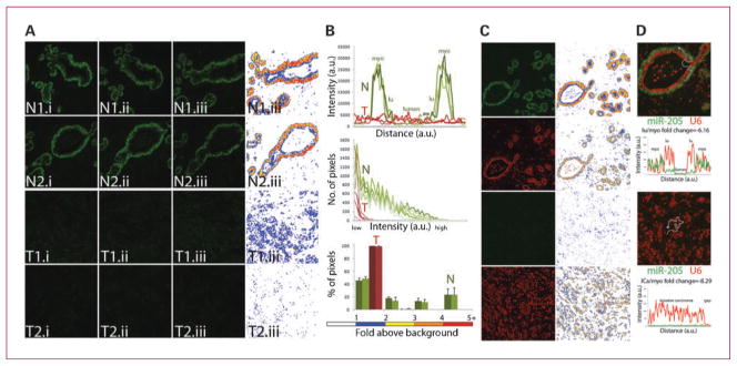
Purpose: High-throughput profiling experiments have linked altered expression of microRNAs (miRNA) to different types of cancer. Tumor tissues are a heterogeneous mixture of not only cancer cells, but also supportive and reactive tumor microenvironment elements. To clarify the clinical significance of altered miRNA expression in solid tumors, we developed a sensitive fluorescence-based in situ hybridization (ISH) method to visualize miRNA accumulation within individual cells in formalin-fixed, paraffin-embedded tissue specimens. This ISH method was implemented to be compatible with routine clinical immunohistochemical (IHC) assays to enable the detection of miRNAs and protein markers in the same tissue section for colocalization and functional studies.

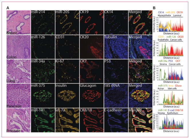
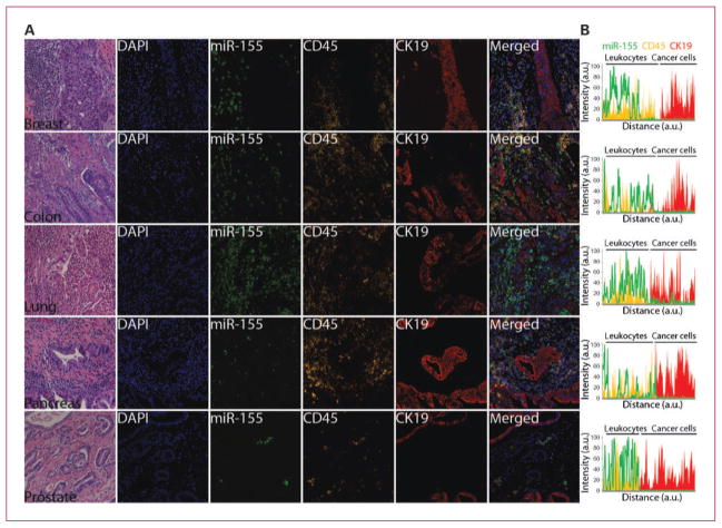
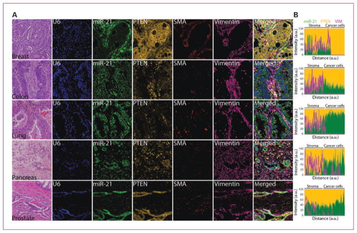
Experimental design: We used this combined ISH/IHC assay to study a subset of cancer-associated miRNAs, including miRNAs frequently detected at low (miR-34a and miR-126) and high (miR-21 and miR-155) levels, in a panel of breast, colorectal, lung, pancreas, and prostate carcinomas.

Results: Despite the distinct histopathologic alterations of each particular cancer type, general trends emerged that pinpointed distinct source cells of altered miRNA expression. Although altered expressions of miR-21 and miR-34a were manifested within cancer cells, those of miR-126 and miR-155 were predominantly confined to endothelial cells and immune cells, respectively. These results suggest a heterogeneous participation of miRNAs in carcinogenesis by intrinsically affecting cancer cell biology or by modulating stromal, vascular, and immune responses.

Conclusions: We described a rapid and sensitive multicolor ISH/IHC assay and showed that it could be broadly applied as an investigational tool to better understand the etiologic relevance of altered miRNA expression in cancer.


Conflict of interest statement

Disclosure of Potential Conflicts of Interest

No potential conflicts of interest were disclosed.

Figures

Fig. 1
Fig. 1
Reproducibility of the ISH method and signal analysis. Matched normal and tumor FFPE breast tissue sections were used to codetect miR-205 and U6 snRNA using FAM2×- and Bio2×-tagged probes, respectively. miR-205 and U6 signals were revealed by sequential TSA reactions with FITC-tyramine (green for miR-205 probe) and rhodamine-tyramine (red for U6 probe) substrates. A, consecutive (i, ii, iii) matched normal (N) and tumor (T) tissue sections were assayed on separate days (1 and 2). Left, raw fluorescent image of the same representative field for each intraexperimental replica. Right, a heat map rendition of intensity classes of miR-205 signal above background noise (see color scale in B). B, the signal intensity of miR-205 was measured across the mammary duct in N1 tissue and across invasive carcinoma lesion in T1 (top). Line profile of N1.iii was slightly shifted to the right (gap) to match the signal from the myoepithelial (myo) and luminal (lu) cellular structures of N1.i and N1.ii. Distribution of pixel intensity of the whole images in A indicate concordant readings for each intraexperimental and interexperimental replica (middle). Columns, mean percentage of pixels within each intensity class for each set of intraexperimental replica; bars, SD (bottom). C, raw images of miR-205 and snRNA U6 were captured with the same exposure and gain setting in normal and tumor tissue (left), which are displayed as a heat map graph (right). D, signal intensity of miR-205 and U6 expression was measured using a line profile tool (merged images are displayed as a 2× magnification of those in C). Background intensity was subtracted from the recorded intensity value and these corrected intensity values were used to generate line graphs. Square dot indicates the beginning of the line profile reading and corresponds with the left-most value of the graph. Average intensity of miR-205 and U6 signal in the indicated structures was used to calculate relative fold decrease of miR-205 expression in luminal and cancer cells compared with myoepithelial cells (lu/myo and ICa/myo, respectively).
Fig. 2
Fig. 2
Codetection of miRNAs with cell type–specific and prognostic protein markers. A, serial FFPE tissue sections of the indicated organs were stained with H&E to reveal histologic features or subjected to ISH assay using FAM2×-tagged probes against miR-34a, miR-126, miR-141, miR-214, and miR-375 mixed with Bio2×-tagged probe against 18S rRNA, BrdU2×-tagged probe against miR-205, or DIG2×-tagged probe against let-7a. miRNA signals were revealed by sequential TSA reactions with FITC-tyramine (green for FAM2× probes), rhodamine-tyramine (orange for BrdU2× and DIG2× probes), and AMCA-tyramine (blue for Bio2× probe) substrates. After HIER as needed (Supplementary Table S2), expression of the indicated proteins was revealed by sequential TSA reactions with Dylight594-tyramine or Dylight680-tyramine (red) and AMCA-tyramine (blue) substrates, each TSA reaction was preceded by incubation with specific antibodies against each protein and appropriate anti-host species/HRP antibody; except for insulin expression, which was revealed by an anti–guinea pig secondary antibody conjugated to Cy3 (orange), obviating the need for the TSA step. B, line profile analysis was used to quantitate the intensity of RNA or protein expression. Background intensity was subtracted and intensity values were normalized setting the point with maximum intensity to 100 and calculating other values in relation to this reference. miRNA expression pattern was plotted as a line; independently, expression patterns of each protein were plotted as stacked areas. Displayed images were modified by optimizing the contrast with the process enhancement function from cellSens software package (Olympus), please see Supplementary Fig. S3 for raw fluorescent images and details on signal analysis.
Fig. 3
Fig. 3
Colocalization of miR-155 with CD45 immune cell marker. A, serial 4-μm FFPE tumor tissue sections of the indicated organs were stained with H&E to reveal histologic features or subjected to standard ISH assays using FAM2×-tagged probe against miR-155. The miR-155 signal was revealed by TSA reaction with FITC-tyramine (green). CK19 expression was revealed by TSA reaction with Dylight680-tyramine (red). After a HIER with citrate, CD45 expression was revealed by TSA reaction with rhodamine-tyramine (orange). Tissue sections were counterstained with nuclear marker 4′,6-diamidino-2-phenylindole (blue). B, line profile analysis was used to quantitate the intensity of RNA or protein expression. Background intensity was subtracted and intensity values were normalized setting the point with maximum intensity to 100, and calculating other values in relation to this reference. miRNA expression pattern was plotted as a line; independently, expression patterns of each protein were plotted as stacked areas. Displayed images were modified by optimizing the contrast with the process enhancement function from cellSens software package (Olympus), please see Supplementary Fig. S4 for raw fluorescent images and details on signal analysis.
Fig. 4
Fig. 4
Codetection of miR-21 with PTEN in clinical specimens. A, serial 4-μm FFPE tumor tissue sections of the indicated organs were stained with H&E to reveal histologic features or subjected to standard ISH assays using FAM2×-tagged probe against miR-21 and Bio2×-tagged probe against snRNA U6. The miR-21 and snRNA U6 signals were revealed by sequential TSA reactions with FITC-tyramine (green) and AMCA-tyramine (blue) substrates. Smooth muscle actin expression (SMA) was revealed by TSA reaction with Dylight594 (red). After a HIER with citrate, PTEN and vimentin expression were revealed by TSA reaction with rhodamine-tyramine (orange) and Dylight680 (magenta), respectively. B, line profile analysis was used to quantitate the intensity of miR-21, PTEN, and vimentin expression. Background intensity was subtracted and intensity values were normalized setting the point with maximum intensity to 100 and calculating other values in relation to this reference. Vimentin expression pattern was plotted as a line; independently, relative expression of miR-21 and PTEN for each data point was plotted as stacked areas. Displayed images were modified by optimizing the contrast with the process enhancement function from cellSens software package (Olympus), please see Supplementary Fig. S5 for raw fluorescent images and details on signal analysis.

References

    1. Lee RC, Ambros V. An extensive class of small RNAs in Caenorhabditis elegans. Science. 2001;294:862–4. - PubMed
    1. Lau NC, Lim LP, Weinstein EG, Bartel DP. An abundant class of tiny RNAs with probable regulatory roles in Caenorhabditis elegans. Science. 2001;294:858–62. - PubMed
    1. Lagos-Quintana M, Rauhut R, Lendeckel W, Tuschl T. Identification of novel genes coding for small expressed RNAs. Science. 2001;294:853–8. - PubMed
    1. Bartel DP. MicroRNAs: target recognition and regulatory functions. Cell. 2009;136:215–33. - PMC - PubMed
    1. Ventura A, Jacks T. MicroRNAs and cancer: short RNAs go a long way. Cell. 2009;136:586–91. - PMC - PubMed

Publication types