. 2010 Aug 17;107(33):14845-50.
doi: 10.1073/pnas.1009874107. Epub 2010 Aug 3.

Distinct subclasses of medium spiny neurons differentially regulate striatal motor behaviors

Affiliations

Distinct subclasses of medium spiny neurons differentially regulate striatal motor behaviors

Helen S Bateup et al. Proc Natl Acad Sci U S A. .

Abstract

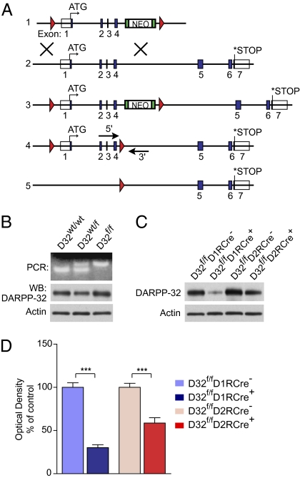
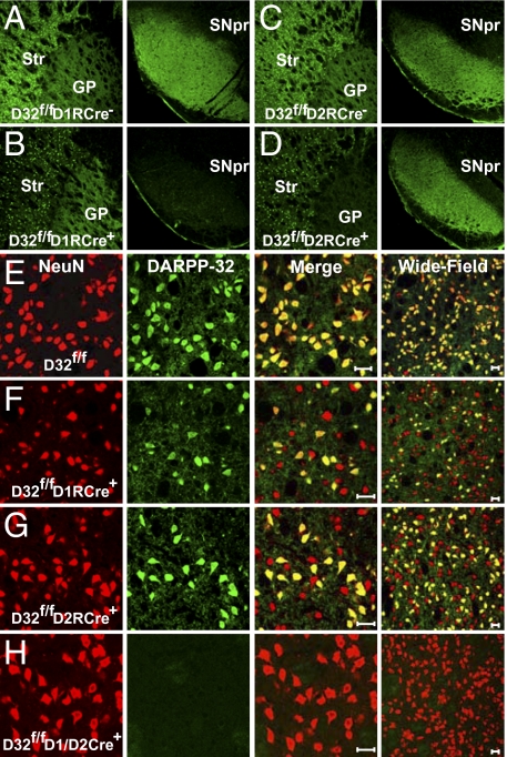
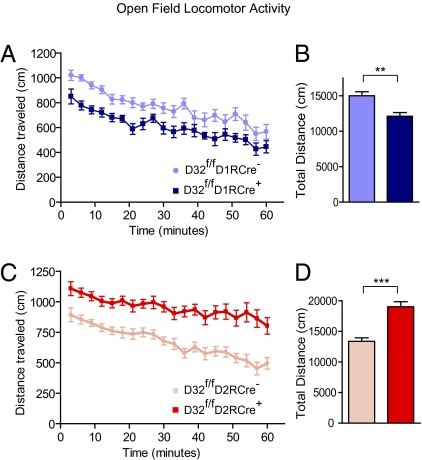
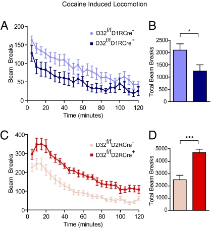
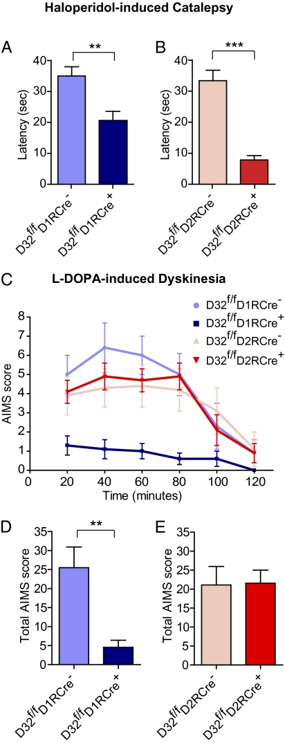
The direct and indirect pathways of the basal ganglia have been proposed to oppositely regulate locomotion and differentially contribute to pathological behaviors. Analysis of the distinct contributions of each pathway to behavior has been a challenge, however, due to the difficulty of selectively investigating the neurons comprising the two pathways using conventional techniques. Here we present two mouse models in which the function of striatonigral or striatopallidal neurons is selectively disrupted due to cell type-specific deletion of the striatal signaling protein dopamine- and cAMP-regulated phosphoprotein Mr 32kDa (DARPP-32). Using these mice, we found that the loss of DARPP-32 in striatonigral neurons decreased basal and cocaine-induced locomotion and abolished dyskinetic behaviors in response to the Parkinson's disease drug L-DOPA. Conversely, the loss of DARPP-32 in striatopallidal neurons produced a robust increase in locomotor activity and a strongly reduced cataleptic response to the antipsychotic drug haloperidol. These findings provide insight into the selective contributions of the direct and indirect pathways to striatal motor behaviors.


Conflict of interest statement

The authors declare no conflict of interest.

Figures

Fig. 1.
Fig. 1.
Generation of DARPP-32 conditional KO mice. (A) Schematic of the generation of the conditional DARPP-32 allele. Blue boxes represent exons; red triangles represent loxP sites. (1) Targeting construct; (2) endogenous DARPP-32 gene; (3) targeted DARPP-32 gene; (4) targeted DARPP-32 gene after in vitro removal of neomycin (NEO) cassette, with arrows showing the location of the genotyping primers; (5) DARPP-32 allele after Cre-mediated deletion of exons 1–4. (B) (Upper) Genotype PCR for the floxed DARPP-32 allele in WT (D32wt/wt), heterozygous (D32wt/f), and homozygous (D32f/f) floxed mice. (Lower) Western blots of striatal lysates from mice from the three genotypes showing equivalent DARPP-32 protein expression and β-actin loading control. (C) Homozygous floxed DARPP-32 mice were bred to D1R-Cre or D2R-Cre mice to conditionally delete DARPP-32. Western blots of striatal lysates from D32f/fD1RCre+ and D32f/fD2RCre+ conditional KO mice show a loss of DARPP-32 protein compared with Cre-negative littermates. (Lower) β-actin loading control. (D) Quantification of striatal DARPP-32 protein levels for mice of the indicated genotypes expressed as a percentage of Cre-negative littermate levels. n = 12 mice per genotype. Bar graphs show group mean ± SEM. ***P < 0.001, unpaired two-tailed t test.
Fig. 2.
Fig. 2.
Selective deletion of DARPP-32 in striatonigral or striatopallidal neurons. (AD) Immunofluoresence performed on brain sections from D32f/fD1RCre (A), D32f/fD1RCre+(B), D32f/fD2RCre (C), and D32f/fD2RCre+ (D) mice using an antibody against DARPP-32. (Left) Somatic DARPP-32 staining in the striatum (Str) and axonal labeling in the GP. (Right) Corresponding axonal DARPP-32 labeling in the SNpr. (E–H) Immunofluoresence of striatal sections from D32f/f (E), D32f/fD1RCre+ (F), D32f/fD2RCre+ (G), and D32f/fD1/D2RCre+ mice (H) costained with antibodies against NeuN (red) and DARPP-32 (green). The third column shows a merged image, and the fourth column shows a merged image of a different brain section at lower magnification. (Scale bar: 20 μm.)
Fig. 3.
Fig. 3.
Corticostriatal LTP is disrupted in DARPP-32 conditional KO mice. (A) LTP was induced by a positive pairing timing protocol as shown at the upper left. Plots show EPSP amplitude as a function of time. The dashed line shows the average EPSP before induction. The induction was performed at the vertical bar. Filled circles show the averages of 12 trials (±SEM). The traces in the upper right show the average EPSP before induction (black trace) and after induction (red trace), from the time periods identified by the horizontal black bars on the plot. Pre, presynaptic; post, postsynaptic. (B and C) D32f/fD1RCre+ and D32f/fD2RCre+ mice were bred to D1R- and D2R-EGFP mice, respectively, to identify D1R and D2R expressing MSNs. EGFP-positive MSNs from D32wt/wtD1RCre+ (B) and D32wt/wtD2RCre+ (C) mice were used as controls and displayed significant LTP. LTP induction was disrupted in EGFP-positive MSNs from D32f/fD1RCre+ mice (B) and D32f/fD2RCre+ mice (C). (D1R MSNs: n = 5 per genotype, P < 0.05; D2R MSNs: n = 5–6 per genotype, P < 0.05, Mann–Whitney rank sum test).
Fig. 4.
Fig. 4.
Basal locomotor activity is altered in DARPP-32 conditional KO mice. Locomotor activity was measured by placing mice individually into an open-field chamber and recording the distance traveled. (A and C) Line graphs showing the distance traveled (in centimeters) in 3-min bins over a 60-min period by mice of the indicated genotypes. (B and D) Bar graphs showing the average total distance traveled (in centimeters) over the 60-min test period. n = 16–25 mice per genotype. Data are expressed as mean ± SEM. **P < 0.01; ***P < 0.001, unpaired two-tailed t test.
Fig. 5.
Fig. 5.
Acute locomotor response to cocaine. Mice were injected with 15 mg/kg of cocaine, and locomotor activity was monitored by photobeams for 120 min. (A and C) Line graphs showing the number of beam breaks plotted versus time for mice of the indicated genotypes. Points represent the number of beam breaks in a 5-min time interval. (B and D) Bar graphs showing the group means ± SEM of total beam breaks for the 120-min test period for mice from each genotype. n = 8–14 mice per genotype. *P < 0.05; ***P < 0.001, unpaired two-tailed t test.
Fig. 6.
Fig. 6.
Differential contribution of striatonigral and striatopallidal neurons to haloperidol-induced catalepsy and L-DOPA–induced dyskinesia. (A and B) Mice were injected with 1.5 mg/kg haloperidol, and 60 min later catalepsy was assessed by measuring the latency until first movement in seconds. Bar graphs show group means ± SEM for mice from the indicated genotypes. n = 15–23 mice per genotype. **P < 0.01; ***P < 0.001, unpaired two-tailed t test. (C and E) Mice received unilateral striatal injections of 6-OHDA and were treated for 10 d with L-DOPA. (C) Time profile of combined axial, limb, orolingual, and locomotive AIMs for mice from the indicated genotypes scored every 20 min over a 120-min period after the last drug administration. (D and E) Bar graphs showing group means ± SEM of total AIMs scored during the observation period for mice of the indicated genotypes. n = 7–9 mice per genotype. **P < 0.01, unpaired two-tailed Student t test.

Similar articles

Cited by

References

    1. Graybiel AM. The basal ganglia: Learning new tricks and loving it. Curr Opin Neurobiol. 2005;15:638–644. - PubMed
    1. Gerfen CR, et al. D1 and D2 dopamine receptor–regulated gene expression of striatonigral and striatopallidal neurons. Science. 1990;250:1429–1432. - PubMed
    1. Albin RL, Young AB, Penney JB. The functional anatomy of basal ganglia disorders. Trends Neurosci. 1989;12:366–375. - PubMed
    1. Gerfen CR. The neostriatal mosaic: Multiple levels of compartmental organization in the basal ganglia. Annu Rev Neurosci. 1992;15:285–320. - PubMed
    1. Miyamoto S, Duncan GE, Marx CE, Lieberman JA. Treatments for schizophrenia: A critical review of pharmacology and mechanisms of action of antipsychotic drugs. Mol Psychiatry. 2005;10:79–104. - PubMed

Publication types

MeSH terms

LinkOut - more resources