. 2010 Sep-Oct;3(9-10):595-604.
doi: 10.1242/dmm.004648. Epub 2010 Aug 3.

Neuroprotection resulting from insufficiency of RANBP2 is associated with the modulation of protein and lipid homeostasis of functionally diverse but linked pathways in response to oxidative stress

Affiliations

Neuroprotection resulting from insufficiency of RANBP2 is associated with the modulation of protein and lipid homeostasis of functionally diverse but linked pathways in response to oxidative stress

Kyoung-in Cho et al. Dis Model Mech. 2010 Sep-Oct.

Abstract

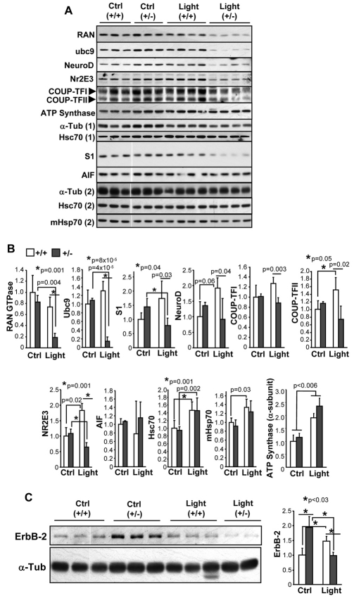
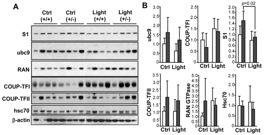
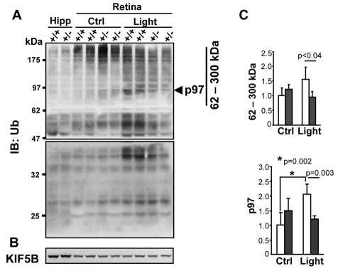
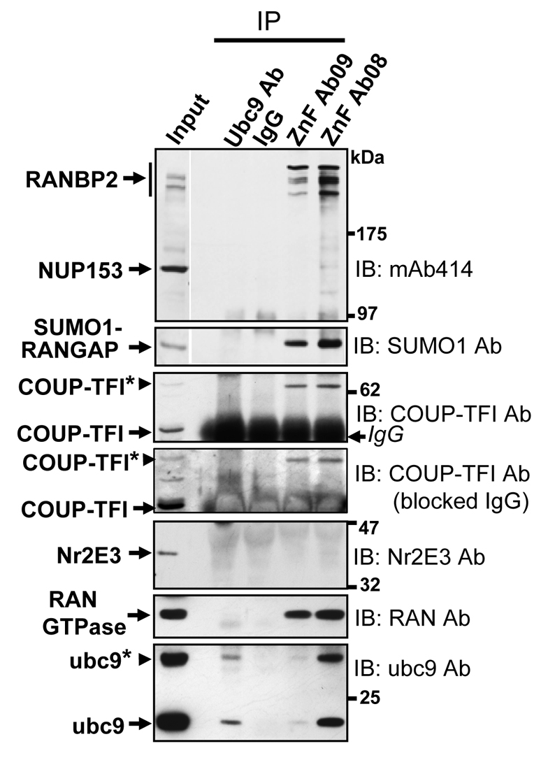
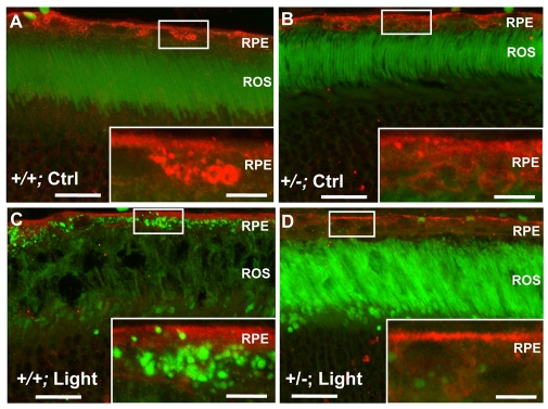
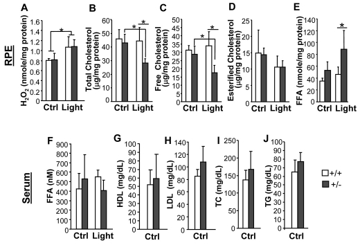
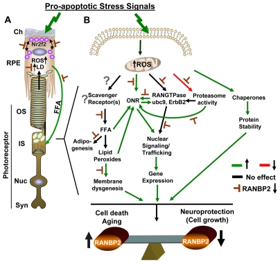
Oxidative stress is a deleterious stressor associated with a plethora of disease and aging manifestations, including neurodegenerative disorders, yet very few factors and mechanisms promoting the neuroprotection of photoreceptor and other neurons against oxidative stress are known. Insufficiency of RAN-binding protein-2 (RANBP2), a large, mosaic protein with pleiotropic functions, suppresses apoptosis of photoreceptor neurons upon aging and light-elicited oxidative stress, and promotes age-dependent tumorigenesis by mechanisms that are not well understood. Here we show that, by downregulating selective partners of RANBP2, such as RAN GTPase, UBC9 and ErbB-2 (HER2; Neu), and blunting the upregulation of a set of orphan nuclear receptors and the light-dependent accumulation of ubiquitylated substrates, light-elicited oxidative stress and Ranbp2 haploinsufficiency have a selective effect on protein homeostasis in the retina. Among the nuclear orphan receptors affected by insufficiency of RANBP2, we identified an isoform of COUP-TFI (Nr2f1) as the only receptor stably co-associating in vivo with RANBP2 and distinct isoforms of UBC9. Strikingly, most changes in proteostasis caused by insufficiency of RANBP2 in the retina are not observed in the supporting tissue, the retinal pigment epithelium (RPE). Instead, insufficiency of RANBP2 in the RPE prominently suppresses the light-dependent accumulation of lipophilic deposits, and it has divergent effects on the accumulation of free cholesterol and free fatty acids despite the genotype-independent increase of light-elicited oxidative stress in this tissue. Thus, the data indicate that insufficiency of RANBP2 results in the cell-type-dependent downregulation of protein and lipid homeostasis, acting on functionally interconnected pathways in response to oxidative stress. These results provide a rationale for the neuroprotection from light damage of photosensory neurons by RANBP2 insufficiency and for the identification of novel therapeutic targets and approaches promoting neuroprotection.


Figures

Fig. 1.
Fig. 1.
Prolonged light exposure and insufficiency of RANBP2 differentially perturb protein homeostasis in retinal neurons. (A) Immunoblot analyses of retinal extracts of 24-week-old wild-type (+/+) and Ranbp2+/− (+/–) mice reared under cyclic (12:12) light conditions (Ctrl) and prolonged light treatment (Light), with antibodies against RAN GTPase, UBC9, NeuroD, Nr2E3, COUP-TFI, COUP-TFII, ATP synthase α subunit, cytosolic Hsc70 (Hsc70), S1 subunit of the 19S cap of the proteasome (S1), mitochondrial apoptosis inducing factor (AIF) and mitochondrial Hsp70 (mHsp70). Acetylated α-tubulin was used as a loading control. (B) Quantification of immunoblots in A by densitometry and normalized against α-tubulin expression. Relative protein levels in wild-type mice under cyclic light conditions were arbitrarily set as 1. In comparison with mice under cyclic (12:12) light conditions (Ctrl), prolonged light exposure (Light) induces a significant decrease in the levels of RAN GTPase, UBC9 and S1 in Ranbp2+/− mice, whereas it causes the upregulation of NeuroD, COUP-TFI and COUP-TFII in wild-type mice. Prolonged light treatment causes an increase and decrease of Nr2E3 levels in wild-type and Ranbp2+/− mice, respectively. No expression change was observed for AIF, whereas chronic light exposure induces an increase in Hsc70, mHsp70 and ATP synthase, regardless of the genotype. (C) Immunoblot (left) and densitometry (right) analyses of ErbB-2 expression (molecular mass ∼185 kDa) in retinal extracts of 24-week-old wild-type (+/+) and Ranbp2+/− (+/−) mice reared under mice under cyclic (12:12) light conditions (Ctrl) and prolonged light treatment (Light). Note that insufficiency of RANBP2 promotes the upregulation of ErbB-2, whereas prolonged light treatment causes a decrease of ErbB-2 levels in both genotypes, but this is more pronounced in Ranbp2+/− mice. Densitometry values are normalized against α-tubulin expression and expressed as arbitrary units as described in B. Densitometry results shown represent the mean ± s.d. of retinas of four mice per group (n=4). Only changes considered significant (P≤0.05) are noted.
Fig. 2.
Fig. 2.
Cyclic and prolonged exposures to light have no effect on the expression levels of several RANBP2 partners in the RPE. (A) Western analyses of RPE extracts of 24-week-old wild-type (+/+) and Ranbp2+/− (+/−) mice reared under cyclic (12:12) light conditions (Ctrl) and prolonged light treatment (Light), with antibodies against proteins employed in Fig. 1. β-Actin was used as loading control. (B) Quantification of immunoblots in A by densitometry and normalized against β-actin expression. Relative protein levels in wild-type (white bars) mice under cyclic light conditions were arbitrarily set as 1. Black bars: Ranbp2+/− mice. With the exception of S1, the expression levels of partners of RANBP2 and cytosolic Hsc70 (Hsc70) did not change significantly under any light conditions. Densitometry values are expressed as arbitrary units. Results shown represent the mean ± s.d. of RPE of four mice per group (n=4). Only changes considered significant (P≤0.05) are noted.
Fig. 3.
Fig. 3.
Insufficiency of RANBP2 suppresses the accumulation of ubiquitylated substrates in response to prolonged light exposure. Immunoblot analyses of retinal and hippocampal (Hipp) extracts of 24-week-old wild-type (+/+) and Ranbp2+/− (+/–) mice reared under cyclic (12:12) light conditions (Ctrl) and prolonged light treatment (Light), with antibodies against ubiquitin (A) and KIF5B (B). In comparison with wild-type mice, a decrease of the levels of ubiquitylated substrates was detected in Ranbp2+/−mice reared under prolonged exposure to light (A). The levels of the RANBP2 partner KIF5B also remained unchanged (B). (C) Quantification by densitometry of ubiquitylated substrates ranging between the apparent molecular masses of 62 and ∼300 kDa [excluding p97, arrowhead (A)], and of a prominent ubiquitylated substrate, p97 [arrowhead, (A)], that are normalized against KIF5B expression. Relative ubiquitin levels in wild-type mice under cyclic light conditions were arbitrarily set as 1. Densitometry values are expressed as arbitrary units. Results shown represent the mean ± s.d. obtained from retinas of four mice per group (n=4) under prolonged light treatment, except for wild-type and Ranbp2+/− mice control groups, where n=7 and n=5, respectively. Only changes considered significant (P≤0.05) are noted. White bars: wild-type mice; black bars: Ranbp2+/− mice.
Fig. 4.
Fig. 4.
RANBP2 selectively associates in vivo with COUP-TFI. Immunoprecipitation of RANBP2 with antibodies against its zinc-finger-rich domain (ZnF Ab09, ZnF Ab08) coprecipitates selectively with a modified isoform of COUP-TFI, but not the homologous nuclear receptor Nr2E3 or an unmodified isoform of COUP-TFI (blocked IgG panel). The RANBP2 partners RAN GTPase, SUMO1-RANGAP and UBC9 also coprecipitate with RANBP2 together with a modified isoform of UBC9 (*). Note the antibody employed against UBC9 is not suitable for immunoprecipitation assays as reflected by the detection of very low levels of self-precipitated UBC9 isoforms. The ZnF antibodies selectively immunoprecipitate RANBP2 (NUP358), but not NUP153 and NUP62. mAb414 is a monoclonal antibody that detects RANBP2, NUP153 and NUP62.
Fig. 5.
Fig. 5.
Prolonged light exposure promotes the RANBP2-dependent accumulation of lipophilic deposits and basal polarization of COUP-TFII in RPE cells. 24-week-old wild-type (+/+) (A,C) and Ranbp2+/− (+/–) (B,D) mice reared under cyclic (12:12) light conditions (Ctrl) (A,B) and prolonged light treatment (Light) (C,D) were stained with the lipophilic dye Bodipy 493/503 for neutral lipids (green) and immunostained with anti-COUP-TFII antibody (red). Note the accumulation of lipophilic deposits in the RPE and swollen RPE cells of wild-type mice (C), whereas COUP-TFII is delocalized towards the basal end of the RPE cells in Ranbp2+/− mice (D). Inset figures are magnified images of RPE areas in squares. RPE, retinal pigment epithelium; ROS, outer segments of rod photoreceptor neurons (rod outer segments). Scale bars, 20 μm; scale bars in inset figures, 5 μm.
Fig. 6.
Fig. 6.
Perturbation of levels of cholesterol and free fatty acids in the RPE in response to gene dosage of Ranbp2 and light-elicited oxidative stress. Prolonged light exposure (Light) induces an increase in H2O2 in the RPE regardless of the genotype (A). In Ranbp2+/− mice, total and free cholesterol levels in the RPE were decreased upon prolonged light exposure (B,C), whereas there was an increase in levels of free fatty acids under the same light conditions (E). The levels of esterified cholesterol in the RPE remain unaltered regardless of the genotype and light conditions (D). Mice of either genotype present no changes in free fatty acids in the serum regardless of the light conditions (F). Wild-type and Ranbp2+/− mice also present no innate changes in high-density lipoprotein cholesterol (HDL) (G), low-density lipoprotein cholesterol (LDL) (H), total cholesterol (TC) (I) and triglycerides (TG) (J) in the serum. Results shown represent the mean ± s.d. of RPE or serum of four mice per group (*, P≤0.05; n=4), except for Ctrl in B, C and D (n=3). Only changes considered significant (P≤0.05) are noted. Ctrl, cyclic (12:12) light treatment; Light, prolonged light treatment. +/+ and +/– are wild-type and Ranbp2+/− mice, respectively.
Fig. 7.
Fig. 7.
Overview of a model depicting the effects of oxidative stress and deficits in RANBP2 in the RPE and photoreceptor neurons. (A) Photoreceptor neurons take up nutrients from the choroidal (Ch) circulation via the RPE cells, which phagocytize the shed membrane-rich outer segments (OS) of photoreceptors and also provide recycled lipid metabolites, such as free fatty acids (FFAs), to photoreceptors by an unknown uptake mechanism. Photo-oxidative stress promotes the accumulation of (neutral) lipid deposits (LDs) probably as a result of impairment of phagocytic and/or proteolytic processes. Orphan nuclear receptors, such as COUP-TFII (Nr2f2), might act as signaling sensors of the homeostasis of lipid metabolites (e.g. FFAs). These effects are affected by insufficiency of RANBP2. (B) In photoreceptors, oxidative stress has multiple effects by causing the upregulation of the expression of orphan nuclear receptors (ONRs) and chaperones (e.g. Hsp70, mHsp70), the downregulation of the proteasome activity and lipid-peroxidation-induced dysgenesis of membranes. Toxic metabolites from lipid peroxidation might also serve as pathophysiological ligands to ONRs. These processes promote a cascade of events culminating in cell death. Insufficiency of RANBP2 counteracts several pathophysiological processes elicited by oxidative stress, it promotes neuroprotection of terminal differentiated photoreceptor neurons to light-elicited oxidative stress and it increases the susceptibility of mitotic cells to uncontrolled cell growth. IS, inner segment compartment of photoreceptor neuron; Nuc, nucleus; OS, outer segment compartment of photoreceptor neuron; ROS, reactive oxygen species; Syn, synapse; green line, stimulatory effect; red line, inhibitory effect; upward arrow, increased level; downward arrow, decreased level. Suppressing effects of RANBP2 are marked by a T-bar.

Similar articles

Cited by

References

    1. Alkuraya FS, Saadi I, Lund JJ, Turbe-Doan A, Morton CC, Maas RL. (2006). SUMO1 haploinsufficiency leads to cleft lip and palate. Science 313, 1751. - PubMed
    1. Andersen JK. (2004). Oxidative stress in neurodegeneration: cause or consequence? Nat Med. 10, S18–S25 - PubMed
    1. Aslanukov A, Bhowmick R, Guruju M, Oswald J, Raz D, Bush RA, Sieving PA, Lu X, Bock CB, Ferreira PA. (2006). RanBP2 modulates Cox11 and Hexokinase I activities and haploinsufficiency of RanBP2 causes deficits in glucose metabolism. PLoS Genet. 2, e177. - PMC - PubMed
    1. Ayaydin F, Dasso M. (2004). Distinct in vivo dynamics of vertebrate SUMO paralogues. Mol Biol Cell 15, 5208–5218 - PMC - PubMed
    1. Bazan NG. (2006). Cell survival matters: docosahexaenoic acid signaling, neuroprotection and photoreceptors. Trends Neurosci. 29, 263–271 - PubMed

Publication types

MeSH terms