. 2010 Oct 8;285(41):31388-98.
doi: 10.1074/jbc.M110.113878. Epub 2010 Aug 3.

Direct binding of the EGF-like domain of neuregulin-1 to integrins ({alpha}v{beta}3 and {alpha}6{beta}4) is involved in neuregulin-1/ErbB signaling

Affiliations

Direct binding of the EGF-like domain of neuregulin-1 to integrins ({alpha}v{beta}3 and {alpha}6{beta}4) is involved in neuregulin-1/ErbB signaling

Katsuaki Ieguchi et al. J Biol Chem. .

Abstract

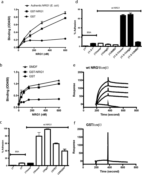
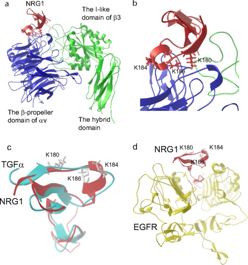
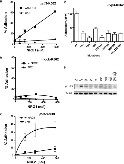
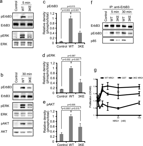
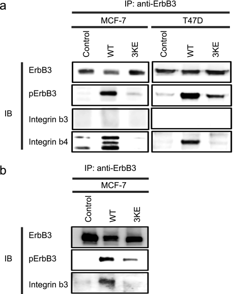
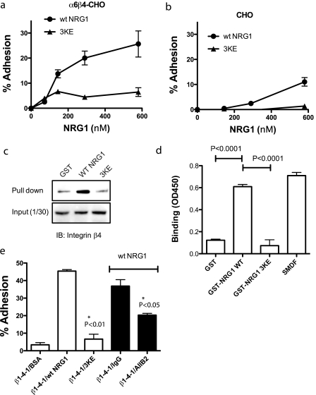
Integrin-growth factor receptor cross-talk plays a role in growth factor signaling, but the specifics are unclear. In a current model, integrins and growth factor receptors independently bind to their ligands (extracellular matrix and growth factors, respectively). We discovered that neuregulin-1 (NRG1), either as an isolated EGF-like domain or as a native multi-domain form, binds to integrins αvβ3 (with a K(D) of 1.36 × 10(-7) m) and α6β4. Docking simulation predicted that three Lys residues at positions 180, 184, and 186 of the EGF-like domain are involved in integrin binding. Mutating these residues to Glu individually or in combination markedly suppressed integrin binding and ErbB3 phosphorylation. Mutating all three Lys residues to Glu (the 3KE mutation) did not affect the ability of NRG1 to bind to ErbB3 but markedly reduced the ability of NRG1 to induce ErbB3 phosphorylation and AKT and Erk1/2 activation in MCF-7 and T47D human breast cancer cells. This suggests that direct integrin binding to NRG1 is critical for NRG1/ErbB signaling. Notably, stimulation of cells with WT NRG1 induced co-precipitation of ErbB3 with α6β4 and with αvβ3 to a much lower extent. This suggests that WT NRG1 induces integrin-NRG1-ErbB3 ternary complex formation. In contrast, the 3KE mutant was much less effective in inducing ternary complex formation than WT NRG1, suggesting that this process depends on the ability of NRG1 to bind to integrins. These results suggest that direct NRG1-integrin interaction mediates integrin-ErbB cross-talk and that α6β4 plays a major role in NRG-ErbB signaling in these cancer cells.


Figures

FIGURE 1.
FIGURE 1.
Direct binding of the EGF-like domain of NRG1 to integrin αvβ3. a and b, the EGF-like domain of NRG1 bound to recombinant soluble αvβ3 in a dose-dependent manner in ELISA-type assays. The EGF-like domain peptide of NRG1 (NRG1α, synthesized in E. coli, R&D systems) (a), SMDF, an isoform of NRG1 (synthesized in eukaryotic cells, R&D systems) (b), GST fusion protein of NRG1, or control GST was immobilized to wells of 96-well microtiter plates. The concentrations of the coating solution are shown. Soluble recombinant integrin αvβ3 (5 μg/ml) was added to the wells in the presence of 1 mm Mn2+ and incubated for 2 h at room temperature. After washing the wells, bound αvβ3 was determined by using anti-β3 antibody and HRP-conjugated anti-mouse IgG. The data are shown as the means ± S.E. of triplicate experiments. c, specific adhesion of CHO cells that express human β3 (β3-CHO) to NRG1 is shown. Wells of 96-wellmicrotiter plate were coated with NRG1 (580 nm) or BSA, and the remaining protein-binding sites were blocked with BSA. Wells were incubated with β3-CHO cells or β1-CHO cells for 1 h at 37 °C in Tyrode's-HEPES buffer with 1 mm MgCl2. Bound cells were quantified. mAb 7E3 (to human β3, 10 μg/ml) and cyclic RGDfV (specific antagonist to αvβ3, 10 μm) blocked the adhesion of β3-CHO cells to WT NRG1. IgG represents purified mouse IgG used as a control. The data are shown as the means ± S.E. of triplicate experiments. d, adhesion of CHO cells that express human β1 (β1-CHO) or β1-3-1 (β1-3-1-CHO) to NRG1 is shown. The β1-3-1 mutation changes the specificity of β1 integrins to that of β3 integrins. Cell adhesion was performed as described above. β1-3-1-CHO cells adhered to WT NRG1, and this binding was blocked by anti-human β1 antibody AIIB2 (10 μg/ml) but not by purified mouse IgG (mIgG). BSA as a control was coated instead of NRG1. The data are shown as the means ± S.E. of triplicate experiments. The results suggest that NRG1 specifically binds to β1-3-1 (as αvβ1-3-1) but not to WT β1 (as αvβ1). e and f, surface plasmon resonance studies of NRG1-αvβ3 interaction are shown. Soluble integrin αvβ3 was immobilized to a sensor chip, and the binding of WT NRG1 and control GST (concentrations at 1000, 500, 250, 125, and 0 nm) was analyzed in the presence of 1 mm MnCl2. KD was calculated as 1.36 × 10⁻⁷ m for WT NRG-1. Control GST did not show significant binding.
FIGURE 2.
FIGURE 2.
Docking simulation of αvβ3-NRG1 interaction. a, a model of NRG1-integrin αvβ3 interaction predicted by docking simulation by using AutoDock3 is shown. The headpiece of integrin αvβ3 (PDB code 1LG5) was used as a target. The model predicts that the EGF-like domain of NRG1 (PDB code 1HAF, blue) binds to the RGD-binding site of the integrin αvβ3 headpiece (green and red). b, the Lys residues at positions 180, 184, and 186 of NRG1α are located at the interface between NRG1 and αvβ3 and were selected for mutagenesis studies. c, superposition of TGFα and NRG1 is shown. d, the Lys residues at positions 180, 184, and 186 of NRG1 are not located in the binding site for EGFR. We replaced TGFα in the TGFα-EGFR complex (PDB code 1MOX) with NRG1 (PDB code 1HAF) by superposing. ErbB3 or ErbB4 is homologous to EGFR.
FIGURE 3.
FIGURE 3.
The 3KE mutant is defective in integrin binding. a and b, the 3KE mutant is defective in binding to αvβ3-K562 cells. Adhesion assays was performed using αvβ3-K562 (a) or mock-transfected cells (b) as described in Fig. 1, except that 1 mm MnCl2 was used. The data are shown as means ± S.E. of triplicate experiments. We obtained similar results using β3-CHO cells. c, the 3KE mutant is defective in binding to β1-3-1 integrin. Adhesion assays was performed using β1-3-1-CHO cells as described above. The data are shown as the means ± S.E. of triplicate experiments. The results suggest that the 3KE mutant is defective in binding to αvβ1-3-1, which mimics αvβ3 in ligand binding. d, mutating the Lys residues alone or in combination suppresses integrin binding. Adhesion assays were performed as described above. We coated wells with GST fusion proteins of NRG1 EGF-like domain (580 nm) and used αvβ3-K562 cells. The numbers represent the positions of Lys residues mutated. e, shown is the signaling function of single and double mutants. We determined the ability of the mutants to induce ErbB3 phosphorylation. MCF-7 cells were serum-starved overnight and stimulated with NRG1(10 nm) for 30 min. Cell lysates were analyzed by Western blotting with antibodies specific to phospho-ErbB3 or ErbB3. Ctl, control.
FIGURE 4.
FIGURE 4.
The 3KE mutant of NRG1 binds to ErbB3. a, binding of the 3KE mutant of NRG1 to recombinant ErbB3 is shown. Recombinant soluble ErbB3 Fc fusion protein (R&D system) was coated onto wells of a 96-well microtiter plate (1 μg/ml). NRG1 WT or 3KE mutant was added to the wells and incubated for 1 h at room temperature. GST was used as a control. After washing the wells, bound GST NRG1 was determined by using anti-GST antibody HRP conjugate. The results suggest that the 3KE mutant of NRG1 binds to ErbB3 at levels nearly comparable with WT NRG1. The data are shown as the means ± S.E. of triplicate experiments. b, shown is a competitive binding assay. Recombinant soluble ErbB3 Fc fusion protein was coated onto wells of 96-well microtiter plate (1 μg/ml). Binding of biotin-labeled NRG1 WT (20 nm) in the presence of increasing concentrations of NRG1 WT, 3KE, or GST is shown. After washing the wells, bound biotin-labeled NRG1 WT was determined by using streptavidin HRP conjugate. The data are shown as the means ± S.E. of triplicate experiments. The results suggest that the 3KE mutant of NRG1 binds to ErbB3 at levels comparable with WT NRG1. c, binding of soluble ErbB3-Fc to immobilized NRG1 is shown. We immobilized NRG1 WT and 3KE proteins to the wells of the 96-well microtiter plate at the indicated coating concentrations and incubated with soluble ErbB3-Fc (1 μg/ml) as described above. Bound ErbB3 was detected using HRP-conjugated anti-His6 tag antibodies and peroxidase substrate. ErbB3-Fc has a His6 tag.
FIGURE 5.
FIGURE 5.
Effect of the 3KE mutation on NRG1 signaling. a and b, the 3KE mutant of NRG1 is defective in inducing ErbB3 phosphorylation, AKT activation, and ERK1/2 activation. MCF-7 cells were serum-starved overnight and stimulated with 10 nm WT and the 3KE mutant of NRG1 for 5 min (a) or 30 min (b). Cell lysates were analyzed by Western blotting. Data are representative of three independent experiments. c–e, levels of phosphorylation were quantified using a luminescence analyzer from triplicate experiments. Data were normalized using WT NRG1 as 1. f, recruitment of p85 of PI3K is shown. Cells were stimulated with WT NRG1 or 3KE, ErbB3 was immuno-purified (IP) from lysates using anti-ErbB3, and immuno-purified materials were analyzed by Western blotting. The p85 subunit of PI3K was detected in lysates of cells stimulated with WT NRG1. Much lower levels of p85 were detected in cells stimulated with 3KE. Data shown are representative of three independent experiments. g, shown is the effect of WT and 3KE NRG1 on the proliferation of MCF-7 cells. Human MCF-7 breast cancer cells were serum-starved overnight and cultured for 48 h with WT or 3KE mutant NRG1. GST was used as a control. Cell number was measured by MTS assays (OD490). The data are shown as the means ± S.E. (n = 3). p < 0.05 by 2-way ANOVA in each case. Data are representative of three independent experiments performed.
FIGURE 6.
FIGURE 6.
WT NRG1 induced co-precipitation of integrin β3 and β4 with ErbB3, whereas 3KE is defective in this function. a, MCF-7 cells were for serum-starved 24 h and stimulated with 10 nm NRG1 WT or 3KE for 5 min. We used 0.7 mg of protein of cell lysate for immunoprecipitation with anti-ErbB3 antibody. Immunoprecipitated materials were analyzed by Western blotting (IB). The levels of ErbB3 phosphorylation were less with the 3KE mutant. Integrin β4 was co-immunoprecipitated with the ErbB3 upon stimulation with WT NRG1, whereas the 3KE mutant was defective in this function. Integrin β3 was not detected under the conditions used. The three bands in co-precipitated β4 in MCF-7 are considered to be (from the top) α6β4 heterodimer, intact β4, and a fragment of β4 based on size. Data are representative of three independent experiments. b, we detected co-precipitation (IP) of β3 with ErbB3 when we used 5× more MCF-7 lysate for co-precipitation experiments using WT NRG1. Data are representative of three independent experiments.
FIGURE 7.
FIGURE 7.
α6β4 binds to WT NRG1, but not to 3KE, through the WPNSDP sequence of β4. WT NRG1 was immobilized to wells of 96-well microtiter plates at 0–580 nm coating concentrations. The remaining protein-binding sites were blocked with BSA. CHO cells that express recombinant α6β4 (α6β4-CHO) (a) or control mock-transfected CHO cells (b) were incubated in the wells for 1 h at 37 °C. Bound cells were measured after gently rinsing the wells. The results suggest that NRG1 binds to integrin α6β4. 3KE NRG1 only weakly interacts with α6β4 (*, p < 0.05.). Data are shown as the means ± S.E. of triplicate experiments. c, pulldown assays are shown. To determine the specificity of binding to α6β4, we incubated GST, GST-WT NRG1, or GST-3KE NRG1 (10 μg) with lysate of α6β4-CHO cells overnight at 4 °C and recovered the material bound to GST proteins using glutathione-Sepharose. We found that WT NRG1 pulled down much more β4 than control GST, and 3KE did not. Data are representative of three independent experiments performed. IB, immunoblot. d, binding of recombinant soluble α6β4 to NRG1 is shown. WT and mutant GST-NRG1 and SMDF were immobilized as described above and incubated with recombinant soluble α6β4 (2 μg/ml), and bound α6β4 was detected using HRP-conjugated anti-Velcro antibody as described under “Experimental Procedures.” e, the β1-4-1 mutant, in which the specificity loop of β1 is replaced with the corresponding amino acid residues of β4, specifically binds to WT NRG1 but not to 3KE. We performed adhesion assays as described above. We coated wells with GST fusion proteins of WT NRG1 and 3KE (580 nm). We used CHO cells that express the β1-4-1 mutant and DMEM for adhesion assays. The data are shown as the means ± S.E. (n = 3).

Similar articles

Cited by

References

    1. Peles E., Bacus S. S., Koski R. A., Lu H. S., Wen D., Ogden S. G., Levy R. B., Yarden Y. (1992) Cell 69, 205–216 - PubMed
    1. Carraway K. L., 3rd, Weber J. L., Unger M. J., Ledesma J., Yu N., Gassmann M., Lai C. (1997) Nature 387, 512–516 - PubMed
    1. Chang H., Riese D. J., 2nd, Gilbert W., Stern D. F., McMahan U. J. (1997) Nature 387, 509–512 - PubMed
    1. Zhang D., Sliwkowski M. X., Mark M., Frantz G., Akita R., Sun Y., Hillan K., Crowley C., Brush J., Godowski P. J. (1997) Proc. Natl. Acad. Sci. U.S.A. 94, 9562–9567 - PMC - PubMed
    1. Falls D. L. (2003) Exp. Cell Res. 284, 14–30 - PubMed

Publication types

MeSH terms