. 2010 Oct 8;285(41):31819-28.
doi: 10.1074/jbc.M110.155846. Epub 2010 Aug 3.

The p38 SAPK is recruited to chromatin via its interaction with transcription factors

Affiliations

The p38 SAPK is recruited to chromatin via its interaction with transcription factors

Isabel Ferreiro et al. J Biol Chem. .

Abstract

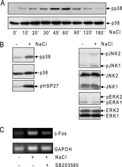
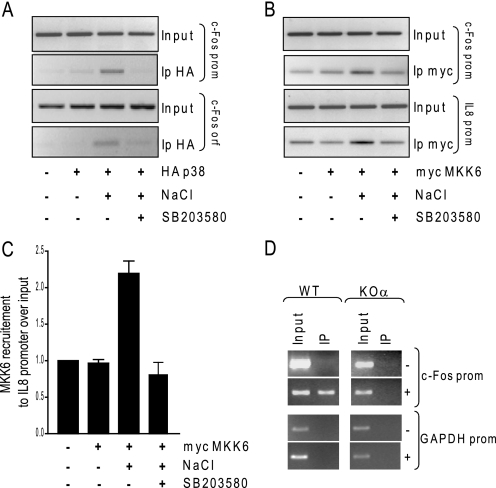
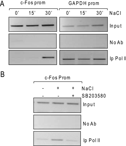
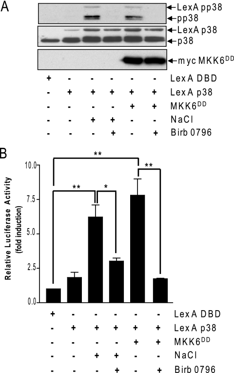
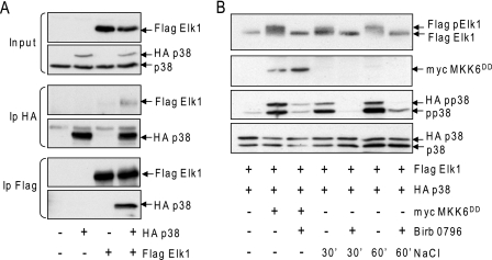
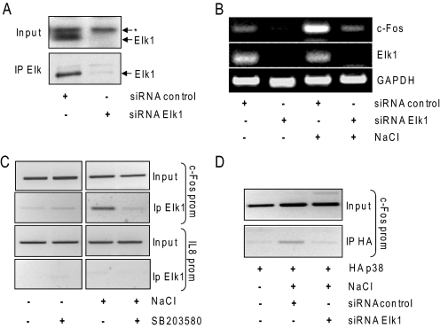
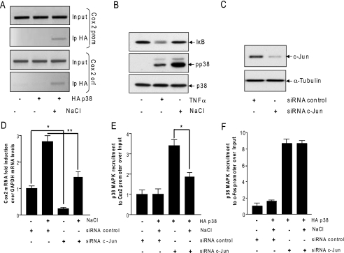
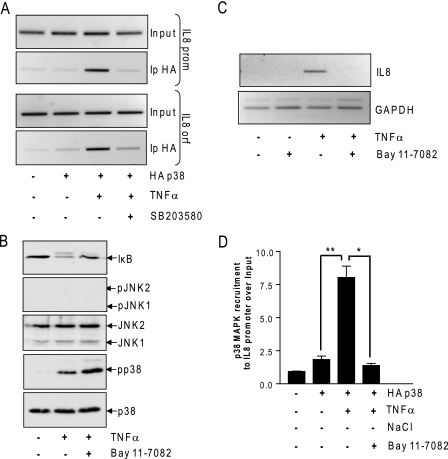
In mammals, the stress-activated protein kinase (SAPK) p38 coordinates a rapid and complex transcriptional program to adapt to sudden changes in the extracellular environment. Although a number of genes have been reported to be under the control of p38, the basic mechanisms of transcriptional regulation by this SAPK remain uncharacterized. Here we show that in response to osmotic shock, anisomycin- or TNFα-activated p38 SAPK is recruited to stress-induced genes. The MAPKK MKK6 is also found at stress-responsive promoters. The recruitment of RNA polymerase II complex to the target promoters requires p38 activity. Moreover, when tethered to DNA as a LexA fusion protein, p38 activates transcription in a stress-regulated manner. Thus, p38 activity allows for recruitment of RNA polymerase and transcription initiation. p38 directly phosphorylates and interacts with the transcription factor Elk1. p38 activity is necessary for the recruitment of Elk1 to the c-Fos promoter, and knocking down Elk1 by siRNAs compromises both p38 recruitment to the c-Fos promoter and c-Fos transcriptional up-regulation upon osmostress. In addition, p38 recruitment to the osmoinducible gene Cox2 and the TNFα target gene IL8 is mediated by the transcription factors AP1 and NFκB, respectively. Therefore, anchoring of active SAPK to target genes is mediated by transcription factors. The presence of active p38 at open reading frames also suggests the involvement of the SAPK in elongation. Taken together, SAPK recruitment to target genes appears to be a broad mechanism to regulate transcription that has been preserved from yeast to mammals.


Figures

FIGURE 1.
FIGURE 1.
Osmostress specifically phosphorylates p38 SAPK and induces c-Fos transcription. A, HeLa cells were stimulated with 100 mm NaCl for the indicated times and analyzed by Western blotting with the indicated antibodies. B, HeLa cells were stimulated with 100 mm NaCl for 45 min and then analyzed by Western blotting with the indicated antibodies. Representative Western blots are shown in A and B. C, HeLa cells were stimulated with 100 mm NaCl for 2 h either in the absence or presence of SB203580. The mRNA levels of c-Fos and GAPDH were analyzed by PCR.
FIGURE 2.
FIGURE 2.
Osmostress induces p38 SAPK and MKK6 recruitment to the c-Fos gene. A, HeLa cells were transfected with pCDNA3–3HA-p38α and treated with 100 mm NaCl for 45 min either in the absence or presence of SB203580. Samples were subjected to ChIP analysis using an anti-HA antibody. B, HeLa cells were transfected with pEFmlinkMKK6 or treated with 100 mm NaCl for 45 min either in the absence or presence of SB203580. ChIP analysis was performed using an anti-Myc antibody. C, relative MKK6DD recruitment to the IL8 promoter. The values represent the mean ± S.D. of three independent experiments performed in triplicate. *, statistically significant (p ≤ 0.05). D, wild type and p38α SAPK knockout MEFs were treated with 100 mm NaCl for 45 min and subjected to ChIP assay using an anti-p38α antibody. Immunoprecipitated DNA fragments were analyzed by PCR.
FIGURE 3.
FIGURE 3.
Osmostress induces RNA Pol II recruitment at the c-Fos promoter. A, HeLa cells were treated with 100 mm NaCl for the indicated times. Binding of RNA Pol II to the c-Fos promoter was determined by ChIP. B, cells were treated with 100 mm NaCl for 30 min and subjected to ChIP analysis using anti-RNA Pol II antibody in the absence or presence of 10 μm SB203580. Immunoprecipitated DNA fragments were analyzed by PCR and ethidium bromide DNA-agarose staining.
FIGURE 4.
FIGURE 4.
Promoter-bound p38 SAPK drives gene transcription. A, HeLa cells were transfected with the shown plasmids. Cells were treated with 100 mm NaCl for 45 min in the absence or presence of BIRB 0796. The inhibitor was added 24 h before harvesting. Lysates were analyzed by Western blotting with the indicated antibodies. Representative Western blots are shown. B, HeLa cells were transfected with the shown plasmids. Cells were treated with 100 mm NaCl in the absence or presence of BIRB 0796 for 24 h before harvesting. The luciferase reporter activity was determined and corrected by the total amount of protein and represented as fold induction over the pCDNA3-LexA-DBD signal, which was considered as 1. The values represent the mean ± S.D. of three independent experiments performed in triplicate. **, statistically significant (p ≤ 0.01) and *, statistically significant (p ≤ 0.05).
FIGURE 5.
FIGURE 5.
p38 SAPK targets and interacts with the transcription factor Elk1. A, HeLa cells were co-transfected with pELK1-Flag and pCDNA3–3HA-p38α. Cell lysates were immunoprecipitated either with anti-HA or anti-Flag antibodies and analyzed by Western blotting with the indicated antibodies. B, HeLa cells were transfected with pELK1-Flag, pCDNA3–3HA-p38α, and pEFmlinkMKK6DD. When indicated, cells were treated with 100 mm NaCl either in the presence or the absence of BIRB 0796. Cleared cell lysates were subsequently analyzed by Western blotting with the indicated antibodies. Representative Western blots are shown in A and B.
FIGURE 6.
FIGURE 6.
p38 SAPK recruitment at the c-Fos promoter requires the transcription factor Elk1. A, HeLa cells were transfected with either control or Elk1 siRNAs and the cell lysates were inmunoprecipitated with anti-Elk1 antibody. Both inputs and immunoprecipitates were analyzed by Western blotting with anti-Elk1 antibody. * marks a nonspecific band. B, HeLa cells were transfected with the indicated siRNAs and treated with 100 mm NaCl for 2 h. The mRNA levels of c-Fos, Elk1, and GAPDH were analyzed by PCR. C, HeLa cells were treated with 100 mm NaCl for 60 min either in the presence or absence SB203580 and subjected to ChIP assay using an anti-Elk1 antibody. D, HeLa cells were transfected with pCDNA3–3HA-p38α along with control or Elk1 siRNAs. Cells were treated with 100 mm NaCl for 60 min and subjected to ChIP assay with anti-HA antibody. Immunoprecipitated DNA fragments were subjected to PCR analysis.
FIGURE 7.
FIGURE 7.
p38 SAPK recruitment to the Cox2 genes requires AP-1 function. A, HeLa cells were transfected with pCDNA3–3HA-p38α, stimulated with 100 mm NaCl for 60 min, and subjected to ChIP assay using an anti-HA antibody. Immunoprecipitated DNA fragments were subjected to PCR. B, HeLa cells were stimulated with 0.1 μg/ml TNFα for 5 min or 100 mm NaCl for 30 min. Cell lysates were subsequently analyzed by Western blotting with the indicated antibodies. C, HeLa cells were transfected with either control or c-Jun siRNAs. Cell lysates were subsequently analyzed by Western blotting with the indicated antibodies. Representative Western blot are shown in B and C. D, HeLa cells were transfected as in C and treated with 100 mm NaCl for 2 h. The mRNA levels of Cox2 were analyzed by real-time PCR, corrected by the GAPDH mRNA levels, and represented as fold induction. E, HeLa cells were co-transfected with pCDNA3–3HA-p38α along with control or c-Jun siRNAs. After the addition of 100 mm NaCl for 60 min, cells were subjected to ChIP analysis with an anti-HA antibody. Immunoprecipitated DNA fragments were analyzed by real-time PCR. Fold induction of p38α SAPK relative enrichment over the input to the Cox2 promoter is shown. Mock transfected cells were taken as 1. F, Hela cells were treated and processed as in E. Fold induction of p38α SAPK relative enrichment over the input to the c-Fos promoter is shown. Means and standard errors of a representative experiment performed in triplicate are shown in D, E, and F. **, statistically significant (p ≤ 0.01) and *, statistically significant (p ≤ 0.05).
FIGURE 8.
FIGURE 8.
p38 SAPK recruitment to the IL8 gene requires NF-κB function. A, HeLa cells were transfected with pCDNA3–3HA-p38α, stimulated with 100 ng/ml TNFα for 60 min in the presence or absence of SB203580 and subjected to ChIP assay using an anti-HA antibody. Immunoprecipitated DNA fragments were subjected to PCR analysis. B, HeLa cells were stimulated with 100 ng/ml TNFα for 5 min. NFκB signaling was blocked by pretreating cells with 50 μm Bay 11-7082 1 h prior to TNFα addition. Cleared cell lysates were subsequently analyzed by Western blotting with the indicated antibodies. Representative Western blots are shown. C, HeLa cells were treated with 100 ng/ml TNFα for 2 h in the absence or presence of 50 μm Bay 11-7082. The mRNA levels of IL8 and GAPDH were analyzed by PCR. D, HeLa cells were transfected with pCDNA3–3HA-p38α and treated with 100 ng/ml TNFα for 45 min or in the absence or presence of 50 μm Bay 11-7082. Cell lysates were subjected to ChIP analysis with an anti-HA antibody. Immunoprecipitated DNA fragments were analyzed by real-time PCR. Fold induction of p38α SAPK relative enrichment over the input is shown. Mock transfected cells were taken as 1. The values represent the mean ± S.D. of two independent experiments performed in triplicate. **, statistically significant (p ≤ 0.01) and *, statistically significant (p ≤ 0.05).

References

    1. Kyriakis J. M., Avruch J. (2001) Physiol. Rev. 81, 807–869 - PubMed
    1. Wagner E. F., Nebreda A. R. (2009) Nat. Rev. Cancer 9, 537–549 - PubMed
    1. Whitmarsh A. J. (2007) Biochim. Biophys. Acta 1773, 1285–1298 - PubMed
    1. Proft M., Pascual-Ahuir A., de Nadal E., Ariño J., Serrano R., Posas F. (2001) EMBO J. 20, 1123–1133 - PMC - PubMed
    1. de Nadal E., Casadomé L., Posas F. (2003) Mol. Cell. Biol. 23, 229–237 - PMC - PubMed

Publication types

MeSH terms

Substances