. 2010 Oct 22;285(43):32878-32887.
doi: 10.1074/jbc.M110.129627. Epub 2010 Aug 3.

The pseudo signal peptide of the corticotropin-releasing factor receptor type 2a decreases receptor expression and prevents Gi-mediated inhibition of adenylyl cyclase activity

Affiliations

The pseudo signal peptide of the corticotropin-releasing factor receptor type 2a decreases receptor expression and prevents Gi-mediated inhibition of adenylyl cyclase activity

Katharina Schulz et al. J Biol Chem. .

Abstract

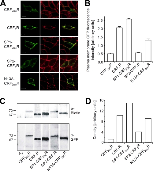
The corticotropin-releasing factor receptor type 2a (CRF(2(a))R) belongs to the family of G protein-coupled receptors. The receptor possesses an N-terminal pseudo signal peptide that is unable to mediate targeting of the nascent chain to the endoplasmic reticulum membrane during early receptor biogenesis. The pseudo signal peptide remains uncleaved and consequently forms an additional hydrophobic receptor domain with unknown function that is unique within the large G protein-coupled receptor protein family. Here, we have analyzed the functional significance of this domain in comparison with the conventional signal peptide of the homologous corticotropin-releasing factor receptor type 1 (CRF(1)R). We show that the presence of the pseudo signal peptide leads to a very low cell surface receptor expression of the CRF(2(a))R in comparison with the CRF(1)R. Moreover, whereas the presence of the pseudo signal peptide did not affect coupling to the G(s) protein, G(i)-mediated inhibition of adenylyl cyclase activity was abolished. The properties mediated by the pseudo signal peptide were entirely transferable to the CRF(1)R in signal peptide exchange experiments. Taken together, our results show that signal peptides do not only influence early protein biogenesis. In the case of the corticotropin-releasing factor receptor subtypes, the use of conventional and pseudo signal peptides have an unexpected influence on signal transduction.


Figures

FIGURE 1.
FIGURE 1.
A, sequence of the uncleaved pseudo signal peptide (SP) of the CRF2(a)R (left) and the conventional cleaved signal peptide of the CRF1R (right). Residue Asn13 of the CRF2(a)R preventing conventional signal peptide functions (18) is depicted in boldface. B, schematic representation of the constructs used in this study. Upper panel, full-length receptor constructs. The signal peptides (SP) and the transmembrane domains (roman numerals) of the CRF2(a)R constructs (black) and the CRF1R constructs (gray) are indicated by boxes. The latin numbers above each construct indicate the number of receptor amino acid residues present (without signal peptide). The latin numbers below each construct indicate the number of amino acids forming the signal peptide. Lower panel, marker protein fusions: N tail sequences fused to GFP are indicated. The latin number above each construct indicate the number of N tail amino acid residues present (without signal peptide). The latin numbers below each construct indicate the number of amino acids forming the signal peptide. H, His tag.
FIGURE 2.
FIGURE 2.
The pseudo signal peptide of the CRF2(a) R maintains its properties in signal peptide exchange experiments. A, localization of the GFP fluorescence signals of the constructs CRF2(a).NT, CRF1.NT, SP1-CRF2(a).NT, SP2-CRF1.NT, and N13A-CRF2(a).NT in transiently transfected HEK 293 cells by confocal LSM. The soluble nonfused GFP protein was used as a control. The horizontal (xy) scans are representative of four independent experiments. Scale bar, 10 μm. n = nucleus. B, analysis of the secretion of the marker protein fusions. Nontransfected cells were used as a control (−). Upper panel, GFP fluorescence measurements. Constructs were purified from the cell culture supernatants of 1.2 × 10⁶ cells, and the GFP fluorescence signals were measured fluorimetrically. Columns represent mean values of three independent experiments each performed in triplicate (S.D.). Lower panel, detection of the purified constructs by immunoblotting using a monoclonal anti-GFP antibody and alkaline phosphatase-conjugated anti-mouse IgG. In each lane, the isolated protein of 2 × 10⁶ cells was loaded. The immunoblot is representative of three independent experiments.
FIGURE 3.
FIGURE 3.
Analysis of signal peptide cleavage of the full-length constructs CRF2(a)R, CRF1R, SP2-CRF1R, SP1-CRF2(a)R, N13A-CRF2(a)R, and the signal peptide deletion mutants ΔSP-CRF2(a)R and ΔSP-CRF1. Receptors were precipitated from transiently transfected HEK 293 cells using an anti-GFP antiserum, digested with PNGaseF to remove all N-glycosylations, and detected by SDS-PAGE immunoblotting using a monoclonal anti-GFP antibody. Nontransfected cells were used as a control for antibody specificity (−). Each lane shows the receptors from 2.5 × 10⁶ cells. The immunoblot is representative of three independent experiments.
FIGURE 4.
FIGURE 4.
The presence of the pseudo signal peptide strongly decreases cell surface receptor expression in transiently transfected HEK 293 cells. A, colocalization of the GFP signals of constructs CRF2(a)R, CRF1R, SP2-CRF1R, SP1-CRF2(a)R, and N13A-CRF2(a)R with the plasma membrane marker trypan blue in live cells by LSM. The GFP signals of the receptor are shown in green (left panels), and trypan blue signals of the cell surface of the same cells are shown in red (center panels). GFP and trypan blue fluorescence signals were computer-overlaid (right panels; overlap is indicated by yellow). The scans show representative cells. Scale bar, 10 μm. Similar data were obtained in three independent experiments. B, quantification of the plasma membrane GFP fluorescence signals of the receptor by automated microscopy. The columns represent mean values of three independent experiments (± S.D.) in arbitrary units. In the individual experiments, the plasma membrane GFP fluorescence intensity of at least 400 cells was analyzed for each construct. C, cell surface biotinylation assay. After labeling of the cell surface receptors with biotin, total receptors were precipitated using an anti-GFP antiserum. Receptors were deglycosylated with PNGaseF to remove all N-glycosylations and detected by SDS-PAGE immunoblotting using a monoclonal anti-biotin antibody (upper part, cell surface receptors) and a monoclonal anti-GFP antibody (lower part; total receptors). Nontransfected cells were used as a control for antibody specificity (−). Each lane shows the receptors from 3.75 × 10⁶ cells. Each immunoblot is representative of three independent experiments. D, densitometric quantification of the protein bands detected by the anti-biotin antibody in C (cell surface receptors).
FIGURE 5.
FIGURE 5.
The pseudo signal peptide increases the amount of immature protein present in the early secretory pathway in transiently transfected HEK 293 cells. A, analysis of the glycosylation state of constructs CRF2(a)R, CRF1R, SP2-CRF1R, and SP1-CRF2(a)R. Receptors were precipitated using a polyclonal anti-GFP antiserum and detected by SDS-PAGE immunoblotting using a monoclonal anti-GFP antibody. Nontransfected cells were used as a control (−). In each lane the same amount of immunoreactive receptor protein was loaded. For each receptor construct, three immunoreactive protein bands are detectable representing the following glycosylation states: mature complex-glycosylated forms (*), immature high mannose forms (§), and immature nonglycosylated forms (#) (see below). The immunoblot is representative of three independent experiments. B, ratio of the individual immunoreactive protein bands for each construct. Intensity of the protein bands was measured densitometrically, and the relative amount was calculated for each construct. Columns represent mean values (± S.D.) of protein band intensity of three independent experiments. C, verification of the identity of the three protein bands for the constructs CRF2(a)R and CRF1R (see also Ref. 18). The receptors were precipitated from transiently transfected HEK 293 cells using a polyclonal anti-GFP antiserum. Samples were treated with EndoH or PNGaseF or left untreated (−). In each lane, the same amount of immunoreactive receptor protein was loaded. Immunoreactive proteins were detected by SDS-PAGE immunoblotting using a monoclonal anti-GFP antibody. Nonglycosylated receptors are EndoH- and PNGaseF-resistant (#); high mannose forms are EndoH- and PNGaseF-sensitive (§); complex-glycosylated receptors are EndoH-resistant and PNGaseF-sensitive (*). The immunoblot is representative of three independent experiments.
FIGURE 6.
FIGURE 6.
The presence of the pseudo signal peptide leads to an increased association of the receptors with the lectin chaperone calnexin. Upper panel, detection of coprecipitated calnexin. Constructs CRF2(a)R, CRF1R, SP2-CRF1R, and SP1-CRF2(a)R were precipitated from transiently transfected HEK 293 cells. Coprecipitated calnexin was detected by SDS-PAGE immunoblotting using a polyclonal anti-calnexin antibody. In each lane, the same amount of immunoreactive receptor protein was loaded to allow comparison of the coprecipitated calnexin. Center panel, loading control. Receptors were detected in the above samples using a monoclonal anti-GFP antibody. All receptors were treated with PNGaseF to remove N-glycosylations allowing comparison of single protein bands. Lower panel, detection of calnexin in total cell lysates using a monoclonal anti-calnexin antibody. In each lane, lysates of 1.25 × 10⁵ cells was loaded. All immunoblots are representative of three independent experiments.
FIGURE 7.
FIGURE 7.
The presence of the pseudo signal peptide prevents Gi-mediated inhibition of adenylyl cyclase activity. A, adenylyl cyclase activity assay using transiently transfected HEK 293 cells expressing constructs CRF2(a)R, CRF1R, SP2-CRF1R, and SP1-CRF2(a)R. Intact cells were stimulated with increasing concentrations of the agonist sauvagine, and a cAMP RIA was performed. Data points represent geometric mean values of three independent experiments, each performed in triplicate. The calculated EC50 values are (95% confidence limits) as follows: CRF2(a)R, 0.12 nm (0.10–0.13); CRF1R (ascending slope), 0.23 nm (0.13–0.41); SP2-CRF1R, 0.27 nm (0.15–0.49); SP1-CRF2(a)R (ascending slope), 0.16 nm (0.15–0.17). B, adenylyl cyclase activity assay using AtT-20 pituitary cells expressing the CRF1R endogenously. The experiment was carried out as described previously in A. Data points represent geometric mean values of three independent experiments, each performed in triplicate. The calculated EC50 value is (95% confidence limits; ascending slope) 6.1 nm (4.3–8.6).
FIGURE 8.
FIGURE 8.
The presence of the pseudo signal peptide prevents Gi-mediated inhibition of adenylyl cyclase activity independent of receptor expression. A, cell surface expression of constructs CRF1R and SP2-CRF1R in various stably transfected HEK 293 cell clones expressing different amounts of the receptors. The GFP fluorescence signals of the receptor were colocalized with the plasma membrane marker trypan blue using a confocal LSM, and their intensity was quantified using an 8-bit grayscale and the LSM software (15). Columns represent mean values of plasma membrane GFP fluorescence intensity of 20 cells (±S.D.). The quantification is representative of three independent experiments. B, flow cytometry quantification of the cell surface receptors of the cell clones A6-CRF1R and B3-SP2-CRF1R. Plasma membrane receptors of 104 cells were quantified using a monoclonal anti-CRF1R antibody directed against the extracellular N tail and a phycoerythrin-conjugated goat anti-mouse IgG. Columns represent mean values of three independent experiments (±S.D.). C, adenylyl cyclase activity assay using the stably transfected HEK 293 cell clones A6-CRF1R and B3-SP2-CRF1R (see A). Intact cells were stimulated with increasing concentrations of the agonist sauvagine, and a cAMP RIA was performed. Data points represent geometric mean values of a single experiment performed in duplicate. Curves are representative of two independent experiments. The calculated EC50 values are (95% confidence limits) as follow: CRF1R (ascending slope), 0.46 nm (0.38–0.56); SP2-CRF1R, 0.48 nm (0.36–0.66).

Similar articles

Cited by

References

    1. Hauger R. L., Risbrough V., Brauns O., Dautzenberg F. M. (2006) CNS Neurol. Disord. Drug Targets 5, 453–479 - PMC - PubMed
    1. Hauger R. L., Grigoriadis D. E., Dallman M. F., Plotsky P. M., Vale W. W., Dautzenberg F. M. (2003) Pharmacol. Rev. 55, 21–26 - PubMed
    1. Denver R. J. (2009) Ann. N.Y. Acad. Sci. 1163, 1–16 - PubMed
    1. Hauger R. L., Risbrough V., Oakley R. H., Olivares-Reyes J. A., Dautzenberg F. M. (2009) Ann. N.Y. Acad. Sci. 1179, 120–143 - PMC - PubMed
    1. Refojo D., Holsboer F. (2009) Ann. N.Y. Acad. Sci. 1179, 106–119 - PubMed

Publication types

MeSH terms

Substances

LinkOut - more resources