. 2010 Dec;24(12):4744-55.
doi: 10.1096/fj.10-166199. Epub 2010 Aug 3.

Kinase-mediated quasi-dimers of EGFR

Affiliations

Kinase-mediated quasi-dimers of EGFR

Erez M Bublil et al. FASEB J. 2010 Dec.

Abstract

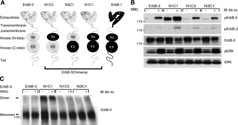
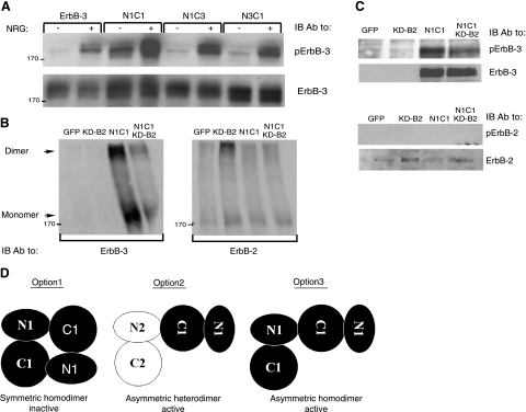
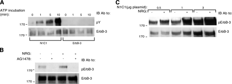
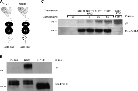
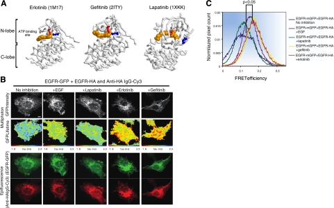
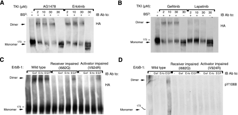
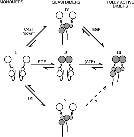
Ligand-induced dimerization of the epidermal growth factor receptor (ErbB-1/EGFR) involves conformational changes that expose an extracellular dimerization interface. Subsequent alterations within the cytoplasmic kinase domain, which culminate in tyrosine phosphorylation, are less understood. Our study addressed this question by using two strategies: a chimeric receptor approach employed ErbB-3, whose defective kinase domain was replaced by the respective part of EGFR. The implanted full-length kinase, unlike its subdomains, conferred dimerization and catalysis. The data infer that the kinase function of EGFR is restrained by the carboxyl tail; once grafted distally to the ectopic tail of ErbB-3, the kinase domain acquires quasi-dimerization and activation. In an attempt to alternatively refold the cytoplasmic tail, our other approach employed kinase inhibitors. Biophysical measurements and covalent cross-linking analyses showed that inhibitors targeting the active conformation of EGFR, in contrast to a compound recognizing the inactive conformation, induce quasi-dimers in a manner similar to the chimeric ErbB-3 molecule. Collectively, these observations unveil kinase domain-mediated quasi-dimers, which are regulated by an autoinhibitory carboxyl tail. On the basis of these observations, we propose that quasi-dimers precede formation of ligand-induced, fully active dimers, which are stabilized by both extracellular and intracellular receptor-receptor interactions.


Figures

Figure 1.
Figure 1.
The N1C1 chimera is constitutively phosphorylated and dimerized. A) Schemes present domain organizations of wild-type ErbB-3 (gray), ErbB-1/EGFR (black), and their chimeric derivatives. N denotes the N lobe and C denotes the C lobe of the respective tyrosine kinase domain. The 4 subdomains of the extracellular regions are denoted I–IV. Dashed line represents the cytoplasmic tail of ErbB-3; solid line represents tail of ErbB-1. Three chimeras are outlined. B) Plasmids encoding ErbB-3 (wild type), N1C1, N1C3, or N3C1 were cotransfected with a plasmid encoding a kinase-dead mutant of ErbB-2 (KD-B2) into CHO cells. After 48 h, cells were stimulated for 5 min without or with NRG (25 ng/ml). Thereafter, cells were detached using a scraper and lysed. Lysates were cleared by centrifugation, resolved by electrophoresis, and then transferred onto a nitrocellulose membrane. Anti-phopsho-ErbB-3 and anti-phospo-ErbB-2 antibodies were used to monitor receptor phosphorylation, whereas an anti-phospho-ERK antibody was used to assess the activation of ERK. An anti-ErbB-3 antibody was used to verify ectopic expression levels, and an antibody against ERK was utilized to verify equal loading. C) Plasmids encoding wild-type ErbB-3, N1C1, N1C3, or N3C1 were cotransfected into CHO cells together with a plasmid encoding the KD-B2 molecule. Cells were stimulated as in B and then washed with cold PBS. Thereafter, cells were detached using a scraper and extracted for 20 min at 4°C in lysis buffer containing the cross-linker BS3 (2 mM). The reaction was terminated by the addition of glycine (20 mM). Lysates were processed as in B. Dimers and monomers (arrows) were detected using an anti-ErbB-3 antibody.
Figure 2.
Figure 2.
The N1C1 chimera incorporates into homodimers rather than heterodimers with ErbB-2. A) Plasmids encoding wild-type ErbB-3, N1C1, N1C3, or N3C1 were transfected into CHO cells. After 48h, cells were stimulated for 5 min without or with NRG (25 ng/ml) and then washed with cold PBS. Thereafter, cells were detached, and their lysates were cleared by centrifugation, resolved by electrophoresis and then transferred onto a nitrocellulose membrane. Anti-phopsho-ErbB-3 and anti-ErbB-3 antibodies were used to monitor phosphorylation or verify expression levels, respectively. B, C) N1C1 and KD-B2 encoding plasmids (3 μg) were transfected into CHO cells, either alone or in combination (each at 1.5 μg). After 48 h, cells were washed with cold PBS, detached using a scraper, and extracted in lysis buffer with (B) or without (C) the cross-linker BS3 (2 mM). The cross-linking reaction was carried out for 20 min at 4°C and terminated by the addition of glycine (20 mM). Thereafter, lysates were cleared by centrifugation, resolved by electrophoresis, and transferred onto a nitrocellulose membrane. presence of ErbB-3 or ErbB-2 in dimers and monomers was monitored using specific antibodies. Phosphotyrosine content of both receptors was examined by using anti-phospho-ErbB-3 or anti-phospho-ErbB-2 antibodies. D) Potential dimerization modes of the N1C1 chimera are indicated. Kinase domains of the N1C1 chimera and ErbB-2 (either KD-B2 or the endogenous receptor) are represented by solid and open shapes, respectively. Option 1 denotes a symmetric inactive homodimer of two N1C1 chimeric receptors, whereas option 2 relates to heterodimerization of the chimeric N1C1 molecule together with the endogenous or the ectopic ErbB-2 molecule. Option 3 indicates an asymmetric active homodimer of two chimeric N1C1 molecules, mediated by their kinase domains.
Figure 3.
Figure 3.
Autophosphorylation of the N1C1 chimera. A) CHO cells were transfected with plasmids encoding ErbB-3 or N1C1. After 48 h, cells were washed with cold PBS and harvested. Cells were lysed for 20 min in lysis buffer (deprived of phosphatase inhibitors), cleared by centrifugation, and then immunoprecipitated using an anti-ErbB-3 antibody. Immunoprecipitates were incubated with a buffer supplemented with ATP (40 μM) for the indicated time intervals. Immunoprecipitates were then washed with cold PBS and resolved by electrophoresis and immunoblotting (IB) utilizing an anti-phosphotyrosine antibody (pY). B) A plasmid encoding the N1C1 chimera was transfected into CHO cells. After 48 h, cells were treated with AG1478 (10 mM) or left untreated for 30 min at 37°C and then stimulated for 5 min with or without NRG (25 ng/ml), and washed with cold PBS. Thereafter, cells were harvested using a scraper and lysed. Lysates were cleared, resolved by electrophoresis, and then transferred onto a nitrocellulose membrane. Phosphorylation and expression of the N1C1 chimera were detected using anti-phospho-ErbB-3 and anti-ErbB-3 antibodies, respectively. C) Cells were transfected as in A with increasing amounts of the N1C1 plasmid and stimulated with or without NRG (25 ng/ml). Thereafter, cells were lysed, and the phosphotyrosine content of the N1C1 chimera was assessed using an anti-phospho-ErbB-3 antibody. ErbB-3 levels were confirmed by stripping and reprobing the membrane with an anti-ErbB-3 antibody.
Figure 4.
Figure 4.
Cytoplasmic tail of ErbB-1/EGFR inhibits basal phosphorylation of the N1C1 chimera. A) Schematic representation of N1C1 in comparison with the N1C1T1 chimera. Note that the chimeras are identical except for the cytoplasmic tails. B) CHO cells were transfected with plasmids encoding the wild-type ErbB-3, N1C1, or the N1C1T1 chimera. At 48 h after transfection, cells were washed with cold PBS, detached, and lysed. Lysates were cleared, resolved by electrophoresis, and then transferred onto a nitrocellulose membrane. Anti-phosphotyrosine and anti-ErbB-3 antibodies (the latter directed against the extracellular region of the receptor) were used to monitor phosphorylation and verify expression levels, respectively. C) CHO cells were transfected with plasmid encoding the N1C1T1 chimera or they were left untreated. After 48 h, cells were stimulated for 5 min with varying concentrations of NRG, and cell lysates were analyzed as in B.
Figure 5.
Figure 5.
FRET/FLIM analysis reveals that erlotinib and gefitinib, but not lapatinib, induce dimerization of ErbB-1/EGFR. A) Three-dimensional structures of the kinase domain of ErbB-1 bound to erlotinib, gefitinib, or lapatinib. Erlotinib (PDB 1M17)- and gefitinib (PDB 2ITY)-bound kinase is active (note salt bridge formed between K721, in red, and E738, in blue, which are in proximity), whereas the lapatinib-bound form (PDB 1XKK) is inactive (K721 and E738 are distant). Structural components of the kinase (N lobe, C lobe, and ATP-binding site) are labeled. B) MCF-7 cells were transfected with vectors encoding EGFR-mGFP and EGFR-HA. Cells were incubated for 24 h, serum starved for 1 h, and stimulated with EGF (50 ng/ml) for 1 h. Alternatively, cells were treated with lapatinib, gefitinib, or erlotinib as indicated (10 μM) for 1 h, prior to fixation and staining with an anti-HA antibody conjugated to Cy-3. Scale bars = 5 μm. C) Cumulative histogram of FRET efficiency between EGFR-mGFP and HA-Cy3 (bound to EGFR-HA) calculated with the following equation in each pixel and averaged per cell: FRET efficiency = 1 tau (da)/tau (control), where tau (da) is the lifetime displayed by cells coexpressing both EGFR-mGFP and EGFR-HA stained with an anti-HA IgG-Cy3, whereas tau (control) is the mean EGFR-mGFP lifetime measurement in the absence of the Cy3 acceptor. Data were obtained from 4–11 cells/treatment group and are representative of 2 independent experiments. Values of P are 0.003 and 0.025, respectively, for comparisons between untreated cells and cells treated with gefitinib or lapatinib, respectively, according to analysis of variance with post hoc testing using Tukey's honest significant test.
Figure 6.
Figure 6.
Cross-linking analysis reveals that AG1478, erlotinib, and gefitinib, but not lapatinib, induce dimerization of ErbB-1/EGFR. A, B) CHO cells were transfected with a plasmid encoding HA-tagged ErbB-1, and 48 h later, they were incubated for 60 min at 37°C with increasing concentrations of AG1478, erlotinib, gefitinib, or lapatinib, as indicated. Cells were then washed with cold PBS and harvested in lysis buffer containing the BS3 cross-linker. After 20 min at 4°C, the cross-linking reaction was terminated by the addition of glycine (20 mM). Thereafter, lysates were cleared by centrifugation, resolved by electrophoresis, and transferred onto a nitrocellulose membrane. Dimers were detected using an antibody against the HA tag. C, D) Cells were transfected with plasmids encoding wild-type ErbB-1/EGFR, a receiver-impaired (I682Q) mutant, or an activator-impaired (V924R) mutant of ErbB-1/EGFR. After 48 h, cells were treated with or without gefitinib (10 μM), erlotinib (10 μM), or EGF (50 ng/ml) and then lysed and cross-linked as in A. Dimers and monomers were detected using an antibody against the HA tag, and tyrosine phosphorylation was monitored using an antibody to the phosphorylated form of EGFR's tyrosine 1068.
Figure 7.
Figure 7.
Proposed mechanism of catalytic activation of ErbB-1/EGFR. EGFR is schematically presented as a large extracellular lobe connected through a transmembrane domain (zigzag line) to a bilobular kinase domain, flanked by a flexible carboxyl tail. Open and shaded lobes symbolize inactive and active conformations. Model assumes that the transition from catalytically inactive monomers (molecular species I) to active dimers (III) involves an intermediate quasi-dimer state (II). Accordingly, in the absence of a ligand EGFR assumes a monomeric inactive conformation (I), in which the cytoplasmic tail inhibits untimely kinase activation. Ligand stimulation enforces conformational changes that stabilize the extracellularly held quasi-dimer (II). Once juxtaposed, the kinase domains displace the carboxyl tails, which results in an active asymmetric dimer held by both extracellular and intracellular interfaces (III). According to the model, tail displacement and subsequent quasi-dimer formation may alternatively initiate with no involvement of the extracellular interface. For example, TKIs that recognize the active conformation of the kinase can extend the tail and form disabled quasi-dimers (V). Alternatively, a mutated terminus, or an ectopic tail (e.g., the tail of N1C1), would enable kinase activation by exposing an interface needed for asymmetric dimer formation (IV). Whether quasi-dimer formation is reversible or influenced by ATP remains unknown.

Similar articles

Cited by

References

    1. Yarden Y., Sliwkowski M. X. (2001) Untangling the ErbB signalling network. Nat. Rev. Mol. Cell. Biol. 2, 127–137 - PubMed
    1. Garrett T. P., McKern N. M., Lou M., Elleman T. C., Adams T. E., Lovrecz G. O., Zhu H. J., Walker F., Frenkel M. J., Hoyne P. A., Jorissen R. N., Nice E. C., Burgess A. W., Ward C. W. (2002) Crystal structure of a truncated epidermal growth factor receptor extracellular domain bound to transforming growth factor alpha. Cell 110, 763–773 - PubMed
    1. Ogiso H., Ishitani R., Nureki O., Fukai S., Yamanaka M., Kim J. H., Saito K., Sakamoto A., Inoue M., Shirouzu M., Yokoyama S. (2002) Crystal structure of the complex of human epidermal growth factor and receptor extracellular domains. Cell 110, 775–787 - PubMed
    1. Hynes N. E., MacDonald G. (2009) ErbB receptors and signaling pathways in cancer. Curr. Opin. Cell Biol. 21, 177–184 - PubMed
    1. Bublil E. M., Yarden Y. (2007) The EGF receptor family: spearheading a merger of signaling and therapeutics. Curr. Opin. Cell Biol. 19, 124–134 - PubMed

Publication types