

Paraneoplastic pemphigus in a burn intensive care unit: case report and review of the literature
- PMID: 20683196
- PMCID: PMC4496247
- DOI: 10.1097/BCR.0b013e3181eed4b4
Paraneoplastic pemphigus in a burn intensive care unit: case report and review of the literature
Abstract
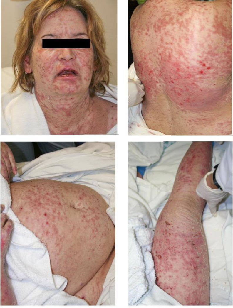
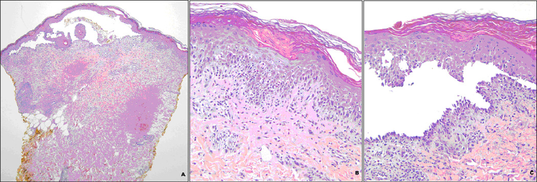
Paraneoplastic pemphigus is a rare cause of acute diffuse blistering in the adult patient. It commonly presents with subepidermal blistering, epidermal necrosis, and symptoms of mucosal irritation, such as conjunctivitis and vaginal ulceration. Because of its rarity, it is frequently misdiagnosed as Stevens-Johnson syndrome or toxic epidermal necrolysis. In this study, the authors will describe clinical and histologic manifestations of paraneoplastic pemphigus. This case report describes a 45-year-old woman with paraneoplastic pemphigus who was admitted and treated in a burn intensive care unit. Although initially diagnosed with Stevens-Johnson syndrome, the patient had progression of desquamation when potentially offending medications were discontinued. Diffuse adenopathy was noted on examination, and biopsy confirmed a low-grade lymphoma. Paraneoplastic pemphigus is a rare but important cause of acute diffuse blistering in adults. This disorder should be considered in the differential diagnosis of patients with diffuse blistering.
Figures
Similar articles
-
Childhood paraneoplastic pemphigus associated with Castleman's tumour.Br J Dermatol. 1997 Jan;136(1):115-7. Br J Dermatol. 1997. PMID: 9039308
-
Case of paraneoplastic pemphigus with follicular lymphoma treated with rituximab.J Dermatol. 2013 Apr;40(4):285-6. doi: 10.1111/1346-8138.12095. Epub 2013 Feb 11. J Dermatol. 2013. PMID: 23398274 No abstract available.
-
Toxic epidermal necrolysis-like presentation of paraneoplastic pemphigus due to underlying thymoma: A clinical conundrum.J Postgrad Med. 2021 Apr-Jun;67(2):119-121. doi: 10.4103/jpgm.JPGM_752_20. J Postgrad Med. 2021. PMID: 33942776 Free PMC article. No abstract available.
-
Paraneoplastic Pemphigus. A Life-Threatening Autoimmune Blistering Disease.Actas Dermosifiliogr. 2017 Dec;108(10):902-910. doi: 10.1016/j.ad.2017.04.024. Epub 2017 Aug 8. Actas Dermosifiliogr. 2017. PMID: 28801011 Review. English, Spanish.
-
Paraneoplastic Pemphigus: Insight into the Autoimmune Pathogenesis, Clinical Features and Therapy.Int J Mol Sci. 2017 Nov 26;18(12):2532. doi: 10.3390/ijms18122532. Int J Mol Sci. 2017. PMID: 29186863 Free PMC article. Review.
Cited by
-
S2k guidelines on the management of paraneoplastic pemphigus/paraneoplastic autoimmune multiorgan syndrome initiated by the European Academy of Dermatology and Venereology (EADV).J Eur Acad Dermatol Venereol. 2023 Jun;37(6):1118-1134. doi: 10.1111/jdv.18931. Epub 2023 Mar 25. J Eur Acad Dermatol Venereol. 2023. PMID: 36965110 Free PMC article.
-
Pemphigus vulgaris mimicking Steven-Johnson syndrome/toxic epidermal necrolysis: report of an unusual case.Dermatol Reports. 2023 May 23;15(3):9649. doi: 10.4081/dr.2023.9649. eCollection 2023 Sep 12. Dermatol Reports. 2023. PMID: 37822989 Free PMC article.
-
Paraneoplastic pemphigus mimicking toxic epidermal necrolysis: An underdiagnosed entity?JAAD Case Rep. 2017 Dec 19;4(1):67-71. doi: 10.1016/j.jdcr.2017.11.002. eCollection 2018 Jan. JAAD Case Rep. 2017. PMID: 29387753 Free PMC article. No abstract available.
-
Paraneoplastic Pemphigus Mimicking Toxic Epidermal Necrolysis Associated with Follicular Lymphoma: Possible Pathological Role of CD8 T Cells.Acta Derm Venereol. 2020 Jul 2;100(14):adv00204. doi: 10.2340/00015555-3557. Acta Derm Venereol. 2020. PMID: 32516424 Free PMC article. No abstract available.
-
Multidisciplinary Management of Paraneoplastic Pemphigus Associated with B-Cell Lymphoma: A Case Report.Int Med Case Rep J. 2025 Jul 8;18:851-857. doi: 10.2147/IMCRJ.S515068. eCollection 2025. Int Med Case Rep J. 2025. PMID: 40656890 Free PMC article.
References
-
- Carr DR, Houshmand E, Heffernan MP. Approach to the acute, generalized, blistering patient. Semin. Cutan. Med. Surg. 2007;26:139–146. - PubMed
-
- Nousari HC, Deterding R, Wojtczack H, Aho S, Uitto J, Hashimoto T, Anhalt GJ. The mechanism of respiratory failure in paraneoplastic pemphigus. N Engl J. Med. 1999 May 6;340(18):1406–1410. - PubMed
-
- Anhalt GJ, Kim SC, Stanley JR, Korman NJ, Jabs DA, Kory M, Izumi H, Ratrie H, 3rd, Mutasim D, Ariss-Abdo L, et al. Paraneoplastic pemphigus. An autoimmune mucocutaneous disease associated with neoplasia. N Engl J. Med. 1990 Dec 20;323(25):1729–1735. - PubMed
-
- Zhu X, Zhang B. Paraneoplastic pemphigus. J Dermatol. 2007 Aug;34(8):503–511. - PubMed
-
- Anhalt GJ. Paraneoplastic pemphigus. J Investig Dermatol Symp Proc. 2004 Jan;9(1):29–33. - PubMed
Publication types
MeSH terms
Substances
Grants and funding
LinkOut - more resources
Full Text Sources
Medical
