

Strategies for vascularization of polymer scaffolds
- PMID: 20683343
- PMCID: PMC2954586
- DOI: 10.231/JIM.0b013e3181f18e38
Strategies for vascularization of polymer scaffolds
Abstract
Biocompatible, degradable polymer scaffolds combined with cells or biological signals are being investigated as alternatives to traditional options for tissue reconstruction and transplantation. These approaches are already in clinical use as engineered tissues that enhance wound healing and skin regeneration. The continued enhancement of these material strategies is highly dependent on the ability to promote rapid and stable neovascularization (new blood vessel formation) within the scaffold. Whereas neovascularization therapies have shown some promise for the treatment of ischemic tissues, vascularization of polymer scaffolds in tissue engineering strategies provides a unique challenge owing to the volume and the complexity of the tissues targeted. In this article, we examine recent advances in research focused on promoting neovascularization in polymer scaffolds for tissue engineering applications. These approaches include the use of growth factors, cells, and novel surgical approaches to both enhance and control the nature of the vascular networks formed. The continued development of these approaches may lead to new tissue engineering strategies for the generation of skin and other tissues or organs.
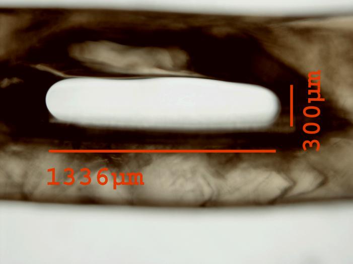
Figures


Similar articles
-
[Progress on strategies to promote vascularization in bone tissue engineering].Zhongguo Gu Shang. 2015 Apr;28(4):383-8. Zhongguo Gu Shang. 2015. PMID: 26072627 Review. Chinese.
-
Vascularization strategies of engineered tissues and their application in cardiac regeneration.Adv Drug Deliv Rev. 2016 Jan 15;96:183-94. doi: 10.1016/j.addr.2015.06.001. Epub 2015 Jun 6. Adv Drug Deliv Rev. 2016. PMID: 26056716 Review.
-
Future Prospects for Scaffolding Methods and Biomaterials in Skin Tissue Engineering: A Review.Int J Mol Sci. 2016 Nov 25;17(12):1974. doi: 10.3390/ijms17121974. Int J Mol Sci. 2016. PMID: 27898014 Free PMC article. Review.
-
Methods for Assessing Scaffold Vascularization In Vivo.Methods Mol Biol. 2019;1993:217-226. doi: 10.1007/978-1-4939-9473-1_17. Methods Mol Biol. 2019. PMID: 31148090
-
Vascularization of engineered tissues: approaches to promote angio-genesis in biomaterials.Curr Top Med Chem. 2008;8(4):300-10. doi: 10.2174/156802608783790983. Curr Top Med Chem. 2008. PMID: 18393893 Free PMC article. Review.
Cited by
-
Regenerative Medicine and Angiogenesis; Challenges and Opportunities.Adv Pharm Bull. 2020 Sep;10(4):490-501. doi: 10.34172/apb.2020.061. Epub 2020 Aug 9. Adv Pharm Bull. 2020. PMID: 33072530 Free PMC article. Review.
-
Non-viral DNA delivery from porous hyaluronic acid hydrogels in mice.Biomaterials. 2014 Jan;35(2):825-35. doi: 10.1016/j.biomaterials.2013.10.014. Biomaterials. 2014. PMID: 24210142 Free PMC article.
-
MMP-sensitive PEG diacrylate hydrogels with spatial variations in matrix properties stimulate directional vascular sprout formation.PLoS One. 2013;8(3):e58897. doi: 10.1371/journal.pone.0058897. Epub 2013 Mar 12. PLoS One. 2013. PMID: 23554954 Free PMC article.
-
Proangiogenic Activity of Endometrial Epithelial and Stromal Cells in Response to Estradiol in Gelatin Hydrogels.Adv Biosyst. 2017 Sep;1(9):1700056. doi: 10.1002/adbi.201700056. Epub 2017 Aug 15. Adv Biosyst. 2017. PMID: 29230433 Free PMC article.
-
Cell-Laden Gradient Hydrogel Scaffolds for Neovascularization of Engineered Tissues.Adv Healthc Mater. 2021 Apr;10(7):e2001706. doi: 10.1002/adhm.202001706. Epub 2021 Jan 29. Adv Healthc Mater. 2021. PMID: 33511790 Free PMC article.
References
-
- MacNeil S. Progress and opportunities for tissue-engineered skin. Nature. 2007;445(7130):874–80. - PubMed
-
- Nikol S, Baumgartner I, Van Belle E, et al. Therapeutic angiogenesis with intramuscular NV1FGF improves amputation-free survival in patients with critical limb ischemia. Mol Ther. 2008;16(5):972–8. - PubMed
-
- Dvorak HF, Detmar M, Claffey KP, et al. Vascular permeability factor/vascular endothelial growth factor: an important mediator of angiogenesis in malignancy and inflammation. Int Arch Allergy Immunol. 1995;107(1–3):233–5. - PubMed
-
- Seliktar D, Zisch AH, Lutolf MP, et al. MMP-2 sensitive, VEGF-bearing bioactive hydrogels for promotion of vascular healing. J Biomed Mater Res A. 2004;68(4):704–16. - PubMed
Publication types
MeSH terms
Substances
Grants and funding
LinkOut - more resources
Full Text Sources
Other Literature Sources
