. 2010 Sep 1;29(17):2979-93.
doi: 10.1038/emboj.2010.164. Epub 2010 Aug 3.

Arabidopsis S6 kinase mutants display chromosome instability and altered RBR1-E2F pathway activity

Affiliations

Arabidopsis S6 kinase mutants display chromosome instability and altered RBR1-E2F pathway activity

Rossana Henriques et al. EMBO J. .

Abstract

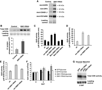
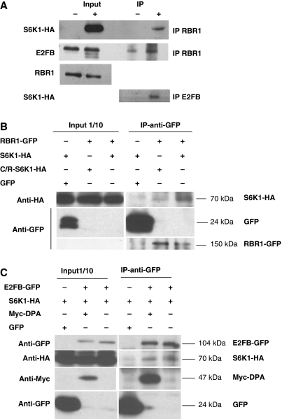
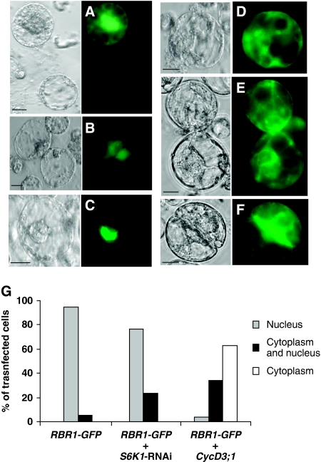
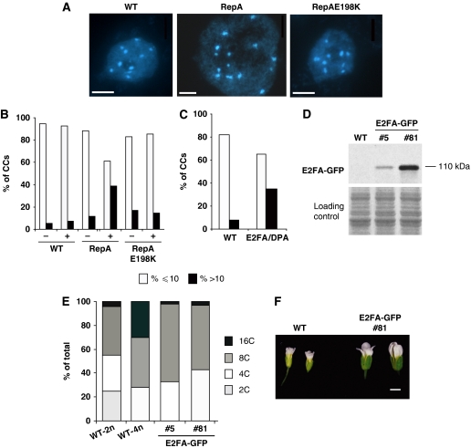
The 40S ribosomal protein S6 kinase (S6K) is a conserved component of signalling pathways controlling growth in eukaryotes. To study S6K function in plants, we isolated single- and double-knockout mutations and RNA-interference (RNAi)-silencing lines in the linked Arabidopsis S6K1 and S6K2 genes. Hemizygous s6k1s6k2/++ mutant and S6K1 RNAi lines show high phenotypic instability with variation in size, increased trichome branching, produce non-viable pollen and high levels of aborted seeds. Analysis of their DNA content by flow cytometry, as well as chromosome counting using DAPI staining and fluorescence in situ hybridization, revealed an increase in ploidy and aneuploidy. In agreement with this data, we found that S6K1 associates with the Retinoblastoma-related 1 (RBR1)-E2FB complex and this is partly mediated by its N-terminal LVxCxE motif. Moreover, the S6K1-RBR1 association regulates RBR1 nuclear localization, as well as E2F-dependent expression of cell cycle genes. Arabidopsis cells grown under nutrient-limiting conditions require S6K for repression of cell proliferation. The data suggest a new function for plant S6K as a repressor of cell proliferation and required for maintenance of chromosome stability and ploidy levels.


Conflict of interest statement

The authors declare that they have no conflict of interest.

Figures

Figure 1
Figure 1
Developmental abnormalities in s6k1s6k2/++ plants. (A) Flowers of WT and s6k1s6k2/++ plants. Scale bar, 1 mm. (B) Anthers from s6k1s6k2/++ and WT plants stained with Alexander dye. Arrows point to pollen. Note the different scale bars, both representing 50 μm. (C) Dissected siliques from WT and s6k1s6k2/++ plants. Arrows point to aborted seeds. Scale bar, 1 mm. (D) Chromosome number in cells of WT leaf epidermis cell, WT pollen meiocyte, s6k1s6k2/++ leaf epidermis cell, s6k1s6k2/++ pollen meiocyte and WT trichomes. Upper row: DAPI staining of chromocentres (CCs) showing a diploid cell (1), meiotic diploid cell in metaphase I (2), tetraploid somatic cell (3), meiotic tetraploid cell in anaphase I (4) and polytenic trichome cell (5). Middle row: FISH with a centromere-specific probe. Lower row: FISH with a specific probe for chromosome 1 pericentromeric regions. Small bars show pairs of chrom.1. A similar increase in chromosome numbers was also found in petal epidermal cells of both s6k1s6k2/++ and s6k1(XVE-RNAi) line 3 plants. Scale bars, 2.5 μm. (E) Flow cytometry measurements of DNA content from flower cells of wild-type (WT-2n), tetraploid wild-type (WT-4n) and s6k1s6k2/++ seedlings. (F) DNA content measurements of leaf no. 1 and 2, 15 DAG. (G) Scanning electron micrographs and corresponding drawings from epidermal leaf surface (third leaf at day 30) of WT and s6k1s6k2/++ plants. Scale bars, 50 μm. (H) Distribution of leaf epidermal cell sizes of s6k1s6k2/++ and WT plants. Variation in cell sizes within classes were analysed from multiple leaf samples and areas (n=299 cells for s6k1s6k2/++ mutants and n=265 cells for WT).
Figure 2
Figure 2
Reducing S6K transcripts in s6k1(XVE-RNAi) plants also leads to ploidy changes. S6K1 (A) and S6K2 (B) transcript levels in s6k1s6k2/++ mutants and the corresponding WT control determined by quantitative RT–PCR. S6K1 (C) and S6K2 (D) transcript levels in s6k1(XVE-RNAi) seedlings (line #3 and #6) and the corresponding WT control in control (−) and 5 μM β-estradiol-treated conditions (+), determined by quantitative RT–PCR. All samples were collected at day 30 after sowing. (E) Percentage of nuclei from leaf epidermal cells having <10 (%<10), 10, and >10 CCs (%>10) from WT (n=59), 5 μM β-estradiol-treated (+) s6k1(XVE-RNAi) line 3 (n=50) plants and s6k1s6k2/++ (n=106) mutants. (F) Percentage of CCs in nuclei from petal epidermal cells in WT (n=166), in 5 μM β-estradiol-treated (+) s6k1(XVE-RNAi) line 3 plants (n=67) and in s6k1s6k2/++ mutants (n=415). (G) Scanning electron micrographs of trichomes from WT and s6k1s6k2/++ leaves. Scale bar, 200 μm. (H) Percentage of trichomes with 3,4,5 and 6 branches in XVE-RNAi empty vector control without β-estradiol (control, −) (n=227), and treated with 5 μM β-estradiol (treated, +) (n=108), s6k1(XVE-RNAi) line 3 control (−) (n=227) and β-estradiol treated (+) (n=292), WT plants (n=279) and s6k1s6k2/++ mutants (n=326). (I) Summary of flow cytometry measurements of DNA content from cells in the first and second leaves at 15 DAG of XVE-RNAi empty vector control and s6k1(XVE-RNAi) constructs.
Figure 3
Figure 3
Cell cycle proteins and CDK activities are upregulated in cells with silenced S6K levels. (A) Detection of E2FB, DPA, CDKB1;1, PSTAIRE-containing CDKA protein levels in mock-transformed (control) Arabidopsis cells, and in cells transformed with the S6K1-RNAi construct, 3 days after transformation (DAT). (B) Total CDK activities of purified CDKs by binding to p13suc1 Sepharose beads from mock (control) and S6K1-RNAi-transformed Arabidopsis cells 3 DAT (triplicates). Upper panel: autoradiogram showing histone H1 phosphorylation by CDKs; lower panel: quantification of the same phosphorylation signal. (C) The CDKB1;1 WT promoter (wt) and CDKB1;1 mutant (Mut) promoter, where the consensus E2F-binding site was mutated (Boudolf et al, 2004) were fused to the GUS-reporter gene and GUS activity measured in mock (control) cells and in cells transformed with S6K1-RNAi and RBR1-RNAi constructs in early stationary stage at 3 DAT. (D) Determination of activity of the RNAR2 WT promoter (wt) fused to the GUS-reporter gene in mock-transformed (control) cells and in cells transformed with S6K1-RNAi and RBR1-RNAi constructs in early stationary stage at 3 DAT. (E) Activity of the CycD3;1 WT promoter (wt) fused to the GUS-reporter gene was determined in similar cells as described in (D). (F) Cell numbers counted in cultures GFP-transformed and cultured in the presence of glucose (GFP/glucose) or mannitol (GFP/mannitol); or transformed with S6K1-RNAi and cultured in presence of glucose (S6K1-RNAi/glucose) or mannitol (S6K1-RNAi/mannitol). Cell numbers were counted at day of transformation (0), and at 2 and 3 DAT. (G) Detection of total CDK activities from the transformed cultures described in (F) at 2 DAT.
Figure 4
Figure 4
S6K1 associates with RBR1 and E2FB. (A) Co-immunoprecipitation of S6K1-HA, E2FB and RBR1. Detection of S6K1-HA in both 1/10th of the input used for immunoprecipitation (IP) of RBR1 and in the RBR1-immunoprecipitate in control cells (−) or in cells where S6K1-HA expression was induced (+) with 5μM of β-estradiol. Detection of E2FB in the same samples of IP-RBR1 (second row). Detection of RBR1 in the input samples used for IP-RBR1 (third row). Detection of S6K1-HA (fourth row) in the E2FB immunoprecipitate (IP-E2FB) from the same input sample as for RBR1-IP. (B) Determination of co-immunoprecipitation of RBR1-GFP with S6K1-HA or C/R-S6K1-HA. Left panel: detection of 1/10th of the input used for IP. Right panel: detection of S6K1-HA, C/R-S6K1-HA, GFP and RBR1-GFP in the RBR1-GFP immunoprecipitate. (C) Determination of co-immunoprecipitation of E2FB-GFP with S6K1-HA or Myc-DPA. Left panel: detection of 1/10th of the input used for IP. Right panel: detection of E2FB-GFP, S6K1-HA, Myc-DPA and GFP in the E2FB-GFP immunoprecipitate.
Figure 5
Figure 5
S6K regulates RBR1 cellular localization. (A) Cells transformed with GFP; (B) S6K1-GFP; (C) RBR1-GFP; (D) RBR1-GFP construct was co-transformed with S6K1-RNAi construct and cultured for 36 h. (E) RBR1-GFP construct was co-transformed with CycD3;1 and cultured for 36 h. (F) Same as in (E) cultured for 72 h. Scale bars, 10 μm. (G) Quantification of the GFP localization signal shown in (CE). RBR1-GFP (n=705 cells); RBR1-GFP+ S6K1-RNAi (n=766 cells); RBR1-GFP+CycD3;1 (n=448 cells) (s.d. values were below 0.04 and are not visible in the graph).
Figure 6
Figure 6
Depletion of RBR1 and over-expression of E2FA/DPA leads to increase in chromocentre (CC) number and polyploidy. (A) DAPI staining of CCs from leaf epidermal cells, showing a diploid cell wild type (WT) (Col-0), a polyploid cell in RepA plants and a diploid cell in mutated RepA198K plants. Scale bars, 1.6 μm. (B) Percentage of nuclei from leaf epidermal cells having <10 or 10 (%⩽10) and >10 CCs (%>10) in WT control (−) (n=372) and in 10 μM dexamethasone-treated (+) (n=379) WT plants; in RepA control (−) (n=305) and in 10 μM dexamethasone-treated (+) (n=407) plants and in RepAE198K control (−) (n=293) and in 10 μM dexamethasone-treated (+) (n=404) plants. (C) Percentage of nuclei from leaf epidermal cells having <10 or 10 (%⩽10) and >10 CCs (%>10) in WT (n=473) and in E2FA/DPA over-expressing lines (n=922). (D) Detection of E2FA-GFP protein level in flowers from the T1 generation of two independent transgenic lines expressing E2FA-GFP under the control of its native promoter. (E) Flow cytometry measurement of the first leaf pair from 15 DAG WT-2n, WT-4n and two T2-independent homozygous E2FA-GFP seedlings from lines described in (D). (F) E2FA-GFP highly expressing line #81 develops larger flowers than WT plants. Scale bar, 1 mm.

Similar articles

Cited by

References

    1. Bernstein KA, Bleichert F, Bean JM, Cross FR, Baserga SJ (2007) Ribosome biogenesis is sensed at the Start cell cycle checkpoint. Mol Biol Cell 18: 953–964 - PMC - PubMed
    1. Bettencourt-Dias M, Giet R, Sinka R, Mazumdar A, Lock WG, Balloux F, Zafiropoulos PJ, Yamaguchi S, Winter S, Carthew RW, Cooper M, Jones D, Frenz L, Glover DM (2004) Genome-wide survey of protein kinases required for cell cycle progression. Nature 432: 980–987 - PubMed
    1. Bögre L, Ökrész L, Henriques R, Anthony RG (2003) Growth signalling pathways in Arabidopsis and the AGC protein kinases. Trends Plant Sci 8: 424–431 - PubMed
    1. Bonatti S, Simili M, Galli A, Bagnato P, Pigullo S, Schiestl RH, Abbondandolo A (1998) Inhibition of the Mr 70 000 S6 kinase pathway by rapamycin results in chromosome malsegregation in yeast and mammalian cells. Chromosoma 107: 498–506 - PubMed
    1. Boudolf V, Vlieghe K, Beemster GTS, Magyar Z, Acosta JAT, Maes S, Van Der Schueren E, Inze D, De Veylder L (2004) The plant-specific cyclin-dependent kinase CDKB1;1 and transcription factor E2Fa-DPa control the balance of mitotically dividing and endoreduplicating cells in Arabidopsis. Plant Cell 16: 2683–2692 - PMC - PubMed

Publication types

MeSH terms