. 2010 Aug;52(2):612-22.
doi: 10.1002/hep.23679.

Human immunodeficiency virus (HIV)-1 infects human hepatic stellate cells and promotes collagen I and monocyte chemoattractant protein-1 expression: implications for the pathogenesis of HIV/hepatitis C virus-induced liver fibrosis

Affiliations

Human immunodeficiency virus (HIV)-1 infects human hepatic stellate cells and promotes collagen I and monocyte chemoattractant protein-1 expression: implications for the pathogenesis of HIV/hepatitis C virus-induced liver fibrosis

Ana C Tuyama et al. Hepatology. 2010 Aug.

Abstract

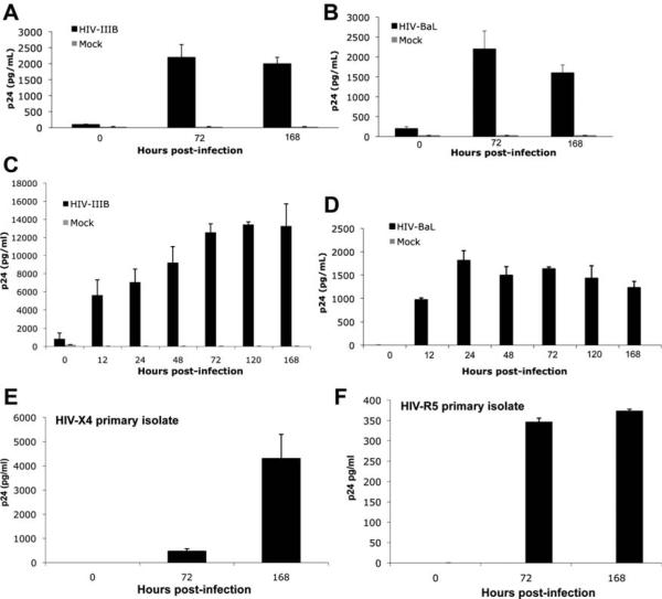
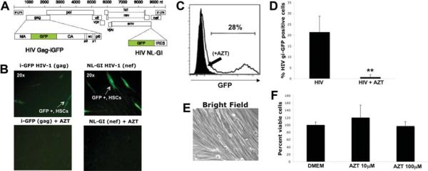
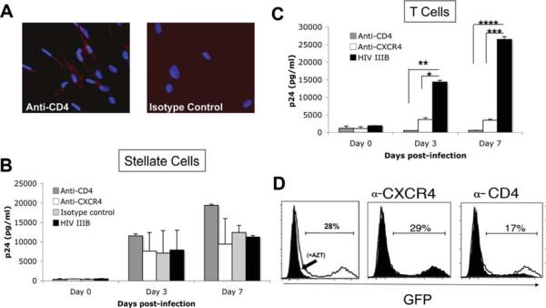
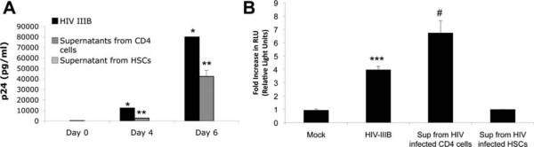
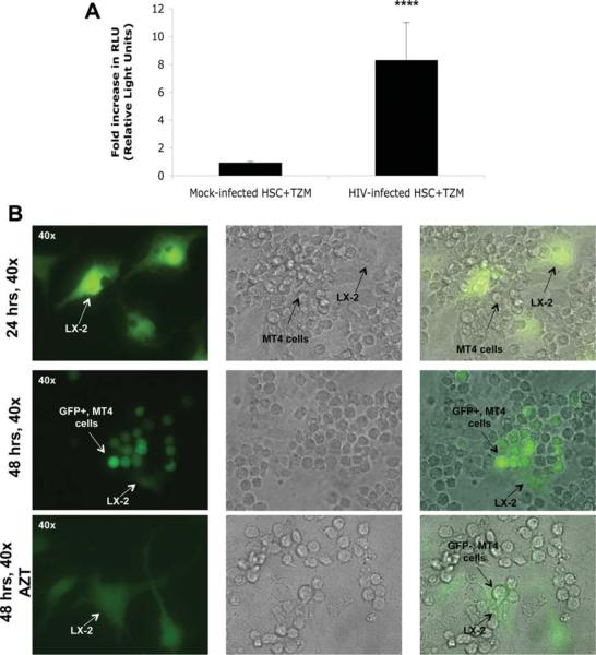
Patients coinfected with human immunodeficiency virus (HIV) and hepatitis C virus (HCV) develop more rapid fibrosis than those infected with HCV only. In HIV/HCV-coinfected patients, fibrosis progression correlates with HIV RNA levels, suggesting a direct role of HIV in liver fibrogenesis. Chemokine (C-C motif) receptor 5 (CCR5) and cysteine-X-cysteine receptor 4 (CXCR4), the two major coreceptors required for HIV entry into cells, are expressed on activated hepatic stellate cells (HSCs), the principle fibrogenic cell type in the liver. We therefore examined whether HIV can infect HSCs, explored the potential mechanisms of viral entry, and assessed the impact of infection as reflected by the ability of HSCs to transfer virus to T lymphocytes and elicit a proinflammatory and profibrogenic response. We report that the laboratory-adapted viruses HIV-IIIB (CXCR4-tropic or X4) and HIV-BaL (CCR5-tropic or R5) and primary HIV isolates can infect both a human stellate cell line, LX-2, and primary human HSCs. HIV entry and gene expression in HSCs was confirmed using HIV-green fluorescent protein (GFP) expression viral constructs in the presence or absence of the reverse-transcriptase inhibitor azidothymidine. CD4 expression on a subset of primary HSCs was demonstrated using fluorescence-activated cell sorting and immunofluorescence staining. Blocking experiments in the presence of anti-CD4, anti-CXCR4, and anti-CCR5 revealed that HIV entry into HSCs is predominantly CD4/chemokine coreceptor-independent. HIV infection promoted HSC collagen I expression and secretion of the proinflammatory cytokine monocyte chemoattractant protein-1. Furthermore, infected LX-2 cells were capable of transferring GFP-expressing virus to T lymphocytes in a coculture system.

Conclusion: Taken together, our results suggest a potential role of HIV in liver fibrosis/inflammation mediated through effects on HSCs. The role of early highly active antiretroviral therapy initiation in patients with HIV/HCV coinfection warrants further investigation.


Figures

Fig. 1
Fig. 1
HSCs are permissive to HIV infection in vitro. LX-2 cells and primary HSCs (passage #3) were infected with HIV-IIIB (X4-tropic), HIV-BaL (R5-tropic), or primary X4-tropic (92UG021) and R5-tropic (92TH007) isolates and culture supernatant collected sequentially for p24 ELISA. Significant p24 was noted up to day #7 after infection in both (A,B) LX-2 and primary (C–F) HSCs. Representative graphs of at least three independent experiments shown. Values are expressed as the mean p24 (pg/mL) ± standard deviation in treated versus mock-infected cells (P < 0.05).
Fig. 2
Fig. 2
Human HSCs support HIV entry and gene expression. (A) HIV-1 Gag-iGFP is an NL4-3-based HIV-1 molecular clone that carries GFP inserted internally into Gag between the MA and CA domains, and HIV-NL-GI (GFP-IRES) is an NL4-3-based HIV-1 molecular clone that carries GFP in place of the nef start codon, with nef expression restored by inserting an internal ribosome entry site as described. Primary HSCs were infected with HIV-1 GFP constructs with or without preincubation with the reverse-transcriptase inhibitor AZT and monitored daily for GFP expression using fluorescence microscopy. (B) GFP expression was noted 48–72 hours after exposure to both X4-tropic HIV NL-GI and HIV Gag-iGFP viral constructs indicating viral entry and both early and late gene expression. Furthermore, preincubation with AZT, a reverse-transcriptase inhibitor, blocked HIV-GFP gene expression in stellate cells (magnification ×40). (C) FACS confirmed a reduction in GFP+ cells in the presence of AZT. (D) Results from three independent experiments are shown graphically. **P < 0.008. (E,F) Lack of toxicity from AZT confirmed by Brightfield Microscopy (E) and MTS assay (F).
Fig. 3
Fig. 3
HIV entry into HSCs is predominantly CD4-independent. (A) Expression of CD4 by primary HSCs was examined by way of immunostaining, revealing a subset of CD4+ cells. A representative image is shown (magnification ×45). (B) To determine whether HIV infection of HSCs is dependent on CD4 and the chemokine coreceptor CXCR4, primary HSCs were preincubated with anti-CD4 (clone Leu 3a, BD Biosciences), anti-CXCR4 (clone 12G5, R&D Systems), or respective isotype control antibodies (all at 10 mg/mL) 30 minutes prior to exposure to the X4-tropic HIV-IIIB, and supernatant was collected for p24 up to 7 days after infection. (C) Efficiency of blocking antibodies was confirmed through simultaneous infection of primary T cells. *P < 0.002. **P < 0.001. ***P < 0.0004. ****P < 0.0006. Representative data from at least three independent experiments performed in triplicate are shown. (D) Primary HSCs were also pretreated with anti-CXCR4 and anti-CD4 antibody prior to exposure to HIV-GI GFP, and FACS for GFP expression performed approximately 3 days after infection. GFP expression was not significantly blocked by anti-CXCR4 or anti-CD4. Although anti-CD4 slightly reduced GFP expression, it was not different than the effect of isotype control. Similarly, infection of HSCs by R5-GFP virus was not blocked by anti-CCR5 antibodies (data not shown). Representative data from three independent experiments are shown.
Fig. 4
Fig. 4
HIV released in culture supernatant from HIV-infected HSCs is noninfectious to TZM cells and CD4 T lymphocytes. Primary HSCs were infected with HIV-IIIB (moi of 0.5) for 4 hours at 37°C, washed extensively, and overlaid with fresh media. (A,B) Culture supernatants collected up to day 7 were incubated with CD4 cells for p24 assay (A) and TZM cells for luciferase activity assay (B). Both purified HIV-IIIB and culture supernatant from infected CD4 cells served as positive controls where increased p24 over time reflects active ongoing replication in CD4 cells (A) (*P < 0.0003, **P < 0.01) and increased luciferase activity in TZM cells reflects activation of the HIV-1 promoter (B) (***P < 0.004, #P < 0.02). No significant p24 or luciferase activity was observed using supernatant derived from infected HSCs. Mock-infected cells served as negative controls.
Fig. 5
Fig. 5
HSCs are able to transfer infectious viral particles to TZM cells and MT4 lymphocytes in coculture systems. (A) For coculture, 5 × 10⁴ TZM cells/well were plated onto 12-well culture plates 1 day prior to infection. HSCs were either mock-infected or infected with HIV-IIIB at an moi of 0.5 for 4 hours at 37°C. Following infection, cells were washed to remove unbound virus, trypsinized, and plated onto TZM cells in a 1:1 ratio. Cells were cocultured for 72 hours, lysed, and analyzed for luciferase activity. An average eight-fold increase in luciferase activity was observed in TZM cells cocultured with HIV-infected HSCs. ****P < 0.0007. Representative data from at least three independent experiments performed in triplicate are shown. (B) LX-2 cells were mock-infected or infected with HIV NL-GI GFP for 24 hours. After viral incubation, the cells were extensively washed and cultured for another 24 hours. Subsequently, the cells were trypsinized and plated in a separate culture plate. After cell attachment, MT4 cells were added to the culture plate in the presence or absence of AZT. Thirty-six to 48 hours after coculture, MT4 cells cocultured with infected LX-2 cells became progressively positive for GFP expression, which was blocked by AZT.
Fig. 6
Fig. 6
HIV promotes HSC collagen I expression and secretion of MCP-1. Primary HSCs (passage #3) were serum-starved for 24 hours, infected with HIV-IIIB for 4 hours at 37°C, washed extensively, and overlaid with fresh media. (A) Western blotting for collagen I was performed on cell lysates prepared 24 and 48 hours after infection. (A) An average 1.5-fold and 2.7-fold increase in collagen I expression was noted at 24 and 48 hours, respectively. β-Tubulin was used as a protein loading control, and fold increase over control is expressed in arbitrary units. (B) Culture supernatant was collected for MCP-1 by way of ELISA. An average 80-fold increase in MCP-1 secretion was observed 48 hours after HSCs were exposed to HIV-IIIB (**P < 0.0004). Representative data from two independent experiments are shown.

Similar articles

Cited by

References

    1. Sherman KE, Rouster SD, Chung RT, Rajicic N. Hepatitis C virus prevalence among patients infected with human immunodeficiency virus: a cross-sectional analysis of the US adult AIDS Clinical Trials Group. Clin Infect Dis. 2002;34:831–837. - PubMed
    1. Thomas D, Astemborski J, Rai R, Anania F, Schaeffer M, Galai N, et al. The natural history of hepatitic C infection: host, viral, and environmental factors. JAMA. 2000;284:450–456. - PubMed
    1. Bonacini M, Govindarajan S, Blatt L, Schmid P, Conrad A, Lindsay K. Patients co-infected with human immunodeficiency virus and hepatitis C virus demonstrate higher levels of hepatic HCV RNA. J Viral Hepatitis. 1999;6:203–208. - PubMed
    1. Benhamou Y, Bochet M, Di Martino V, Charlotte F, Azria F, Coutellier A, et al. Liver fibrosis progression in human immunodeficiency virus and hepatitis C virus coinfected patients. The Multivirc Group. HEPATOLOGY. 1999;30:1054–1058. - PubMed
    1. Macias J, Berenguer J, Japon M, Giron J, Rivero A, Lopez-Cortes L, et al. Fast fibrosis progression between repeated liver biopsies in patients coinfected with human immunodeficiency virus/hepatitis C virus. HEPATOLOGY. 2009;50:1056–1063. - PubMed