Review
. 2010 Dec;152A(12):2962-73.
doi: 10.1002/ajmg.a.33568.

Regulation of facial morphogenesis by endothelin signaling: insights from mice and fish

Affiliations
Review

Regulation of facial morphogenesis by endothelin signaling: insights from mice and fish

David E Clouthier et al. Am J Med Genet A. 2010 Dec.

Abstract

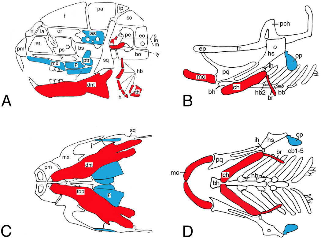
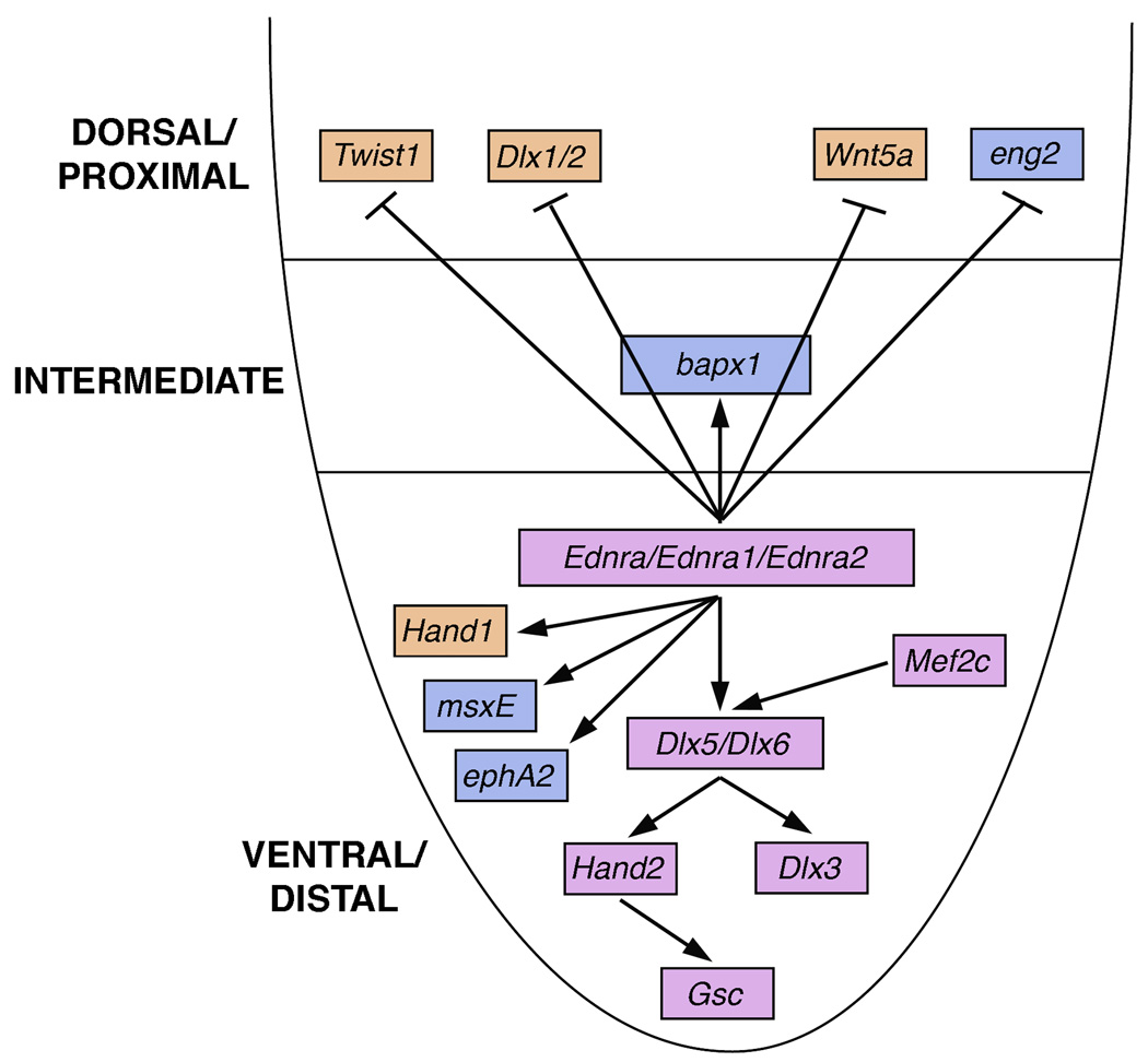
Craniofacial morphogenesis is accomplished through a complex set of developmental events, most of which are initiated in neural crest cells within the pharyngeal arches. Local patterning cues from the surrounding environment induce gene expression within neural crest cells, leading to formation of a diverse set of skeletal elements. Endothelin-1 (Edn1) is one of the primary signals that establishes the identity of neural crest cells within the mandibular portion of the first pharyngeal arch. Signaling through its cognate receptor, the endothelin-A receptor, is critical for patterning the ventral/distal portion of the arch (lower jaw) and also participates with Hox genes in patterning more posterior arches. Edn1/Ednra signaling is highly conserved between mouse and zebrafish, and genetic analyses in these two species have provided complementary insights into the patterning cues responsible for establishing the craniofacial complex as well as the genetic basis of facial birth defect syndromes.


Figures

Figure 1
Figure 1
Pharyngeal arch morphology in mouse and zebrafish. Lateral (A, B) and ventral (C, D) views of the craniofacial skeletons of embryonic day (E)18.5 (mouse) and 5 days postfertilization larvae (dpf) (fish). Within the mandibular and hyoid arch skeletons, Edn1 signaling is required for distal (ventral) arch cartilage and bone (red) and inhibits formation of more proximal (dorsal) cartilage and bone (blue). as, alisphenoid; bb, basibranchial; bh, basihyal; bo, basioccipital; br, branchiostegal ray; bs, basisphenoid; cb, ceratobranchial; ch, ceratohyal; dnt, dentary; eo, exoccipital; ep, ethmoid plate; et, ethmoid; f, frontal; h, hyoid; hb, hypobranchial; hs, hyosymplectic; ih, interhyal; in, incus; ip, interparietal; j, jugal; la, lacrimal; m, malleus; mc, Meckel’s; mx, maxilla; n, nasal; op, opercle; or, orbital; p, palatine; pa, parietal; pch, prechordal; pe, petrosal; pl, palatine; pm, premaxilla; pq, palatoquadrate; ps, presphenoid; ptr, pterygoid; s, stapes; so, supraoccipital; sq, squamosal; tbp, trabecular basal plate; th, thyroid; tr, trabecula; ty, tympanic ring; v, vomer.
Figure 2
Figure 2
Intracellular signaling pathway within cranial neural crest cells mediated by endothelin-1 (Edn1). Preproendothelin-1 coming from cells within the pharyngeal arch environment undergoes two processing events (Furin A/B and endothelin converting enzyme 1/2 (Ece1, 2) before binding to the endothelin-A receptor (Ednra). This initiates signaling through Gαq/Gα11, resulting in phospholipase-β3 activity (Plcβ3) and thus changes in gene expression. Genetic evidence that individual Edn1/Ednra pathway members regulate craniofacial development has come from either mouse (highlighted in orange), zebrafish (highlighted in blue) or both (highlighted in orange/blue).
Figure 3
Figure 3
Schematic of Edn1/Ednra signaling with the mandibular portion of the first pharyngeal arch. Ednra (Ednra1/2 in zebrafish) signaling leads to the induction of gene expression in the ventral (distal) and intermediate domains of the mandibular arch while repressing the expression of dorsal (proximal) genes in the intermediate and ventral arch domains. Many factors induced or repressed by Ednra signaling are conserved between mouse and zebrafish (highlighted in purple), though there are apparent species-specific differences between mouse (highlighted in orange) and zebrafish (highlighted in blue).

Similar articles

Cited by

References

    1. Abe M, Ruest L-B, Clouthier DE. Fate of cranial neural crest cells during craniofacial development in endothelin-A receptor deficient mice. Int J Dev Biol. 2007:51. - PMC - PubMed
    1. Arnold MA, Kim Y, Czubryt MP, Phan D, McAnally J, Qi X, Shelton JM, Richardson JA, Bassel-Duby R, Olson EN. MEF2C transcription factor controls chondrocyte hypertrophy and bone development. Dev Cell. 2007;12:377–389. - PubMed
    1. Barbosa AC, Funato N, Chapman S, McKee MD, Richardson JA, Olson EN, Yanagisawa H. Hand transcription factors cooperatively regulate development of the distal midline mesenchyme. Dev Biol. 2007;310:154–168. - PMC - PubMed
    1. Barlow A, de Graaff E, Pachnis V. Enteric nervous system progenitors are coordinately controlled by the G protein-coupled receptor EDNRB and the receptor tyrosine kinase RET. Neuron. 2003;40:905–916. - PubMed
    1. Baynash AG, Hosoda K, Giaid A, Richardson JA, Emoto N, Hammer RE, Yanagisawa M. Interaction of endothelin-3 with endothelin-B receptor is essential for development of epidermal melanocytes and enteric neurons. Cell. 1994;79:1277–1285. - PubMed

Publication types

MeSH terms