. 2010 Sep;42(9):1525-35.
doi: 10.1016/j.biocel.2010.05.011.

Interaction of ibogaine with human alpha3beta4-nicotinic acetylcholine receptors in different conformational states

Affiliations

Interaction of ibogaine with human alpha3beta4-nicotinic acetylcholine receptors in different conformational states

Hugo R Arias et al. Int J Biochem Cell Biol. 2010 Sep.

Abstract

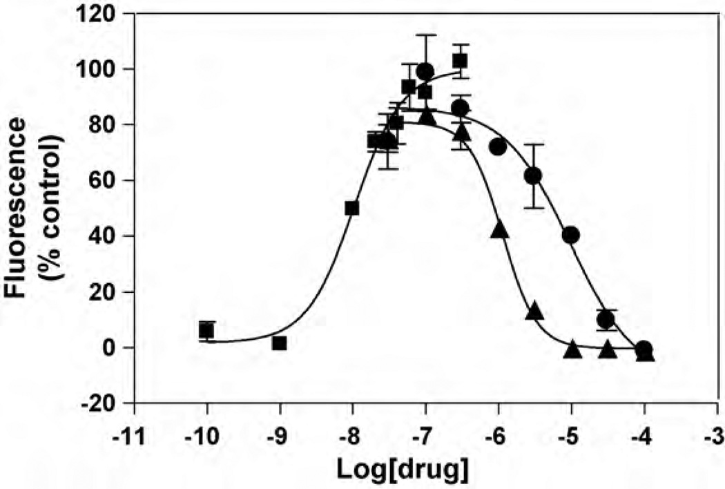
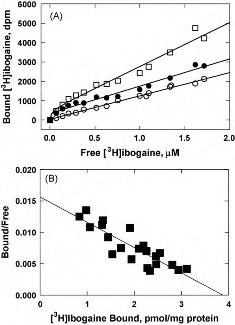
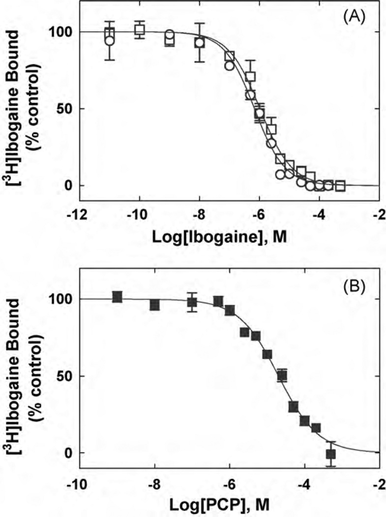
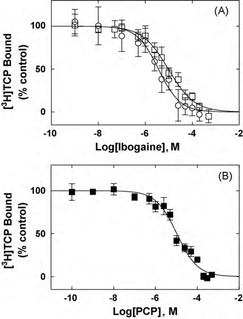
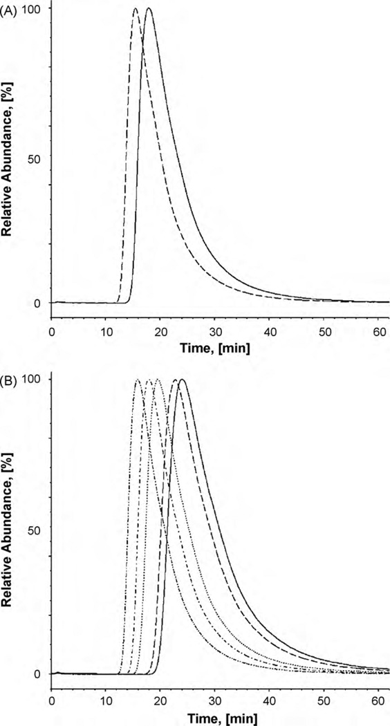
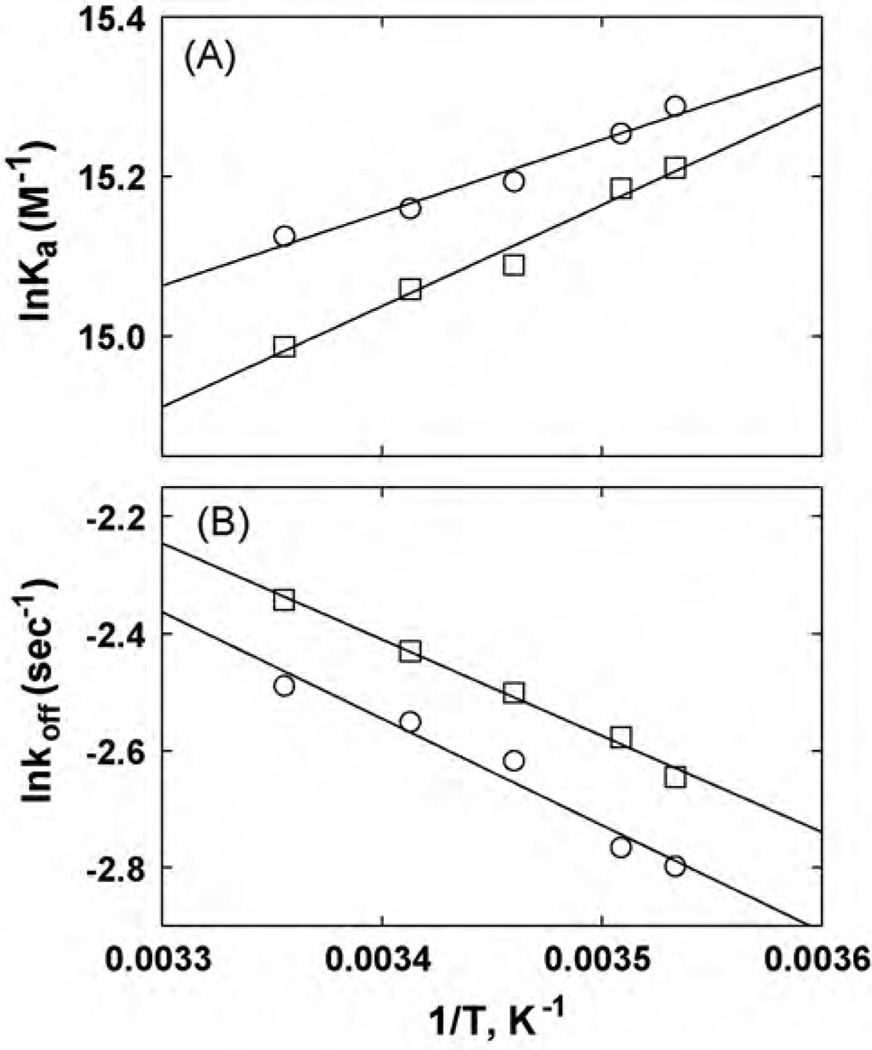
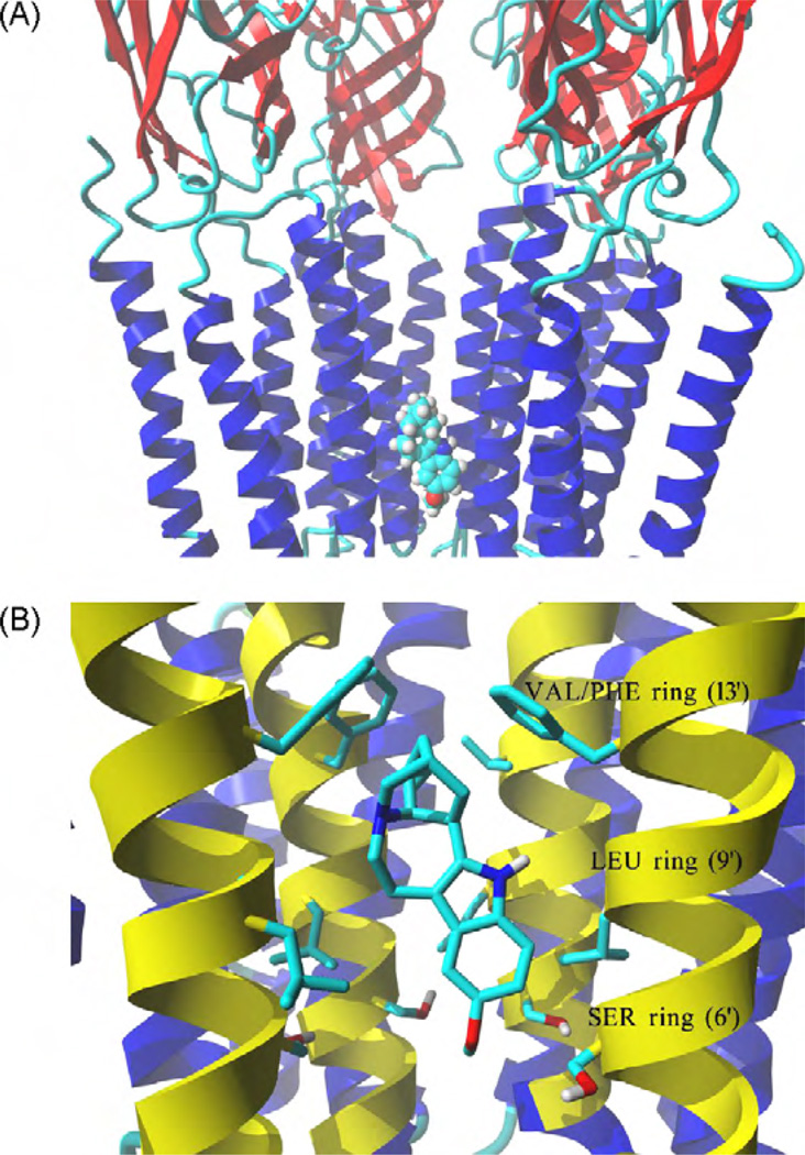
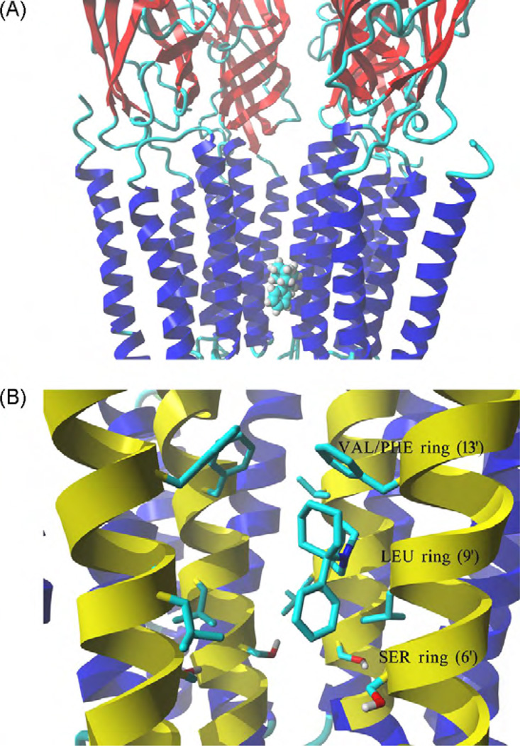
The interaction of ibogaine and phencyclidine (PCP) with human (h) alpha3beta4-nicotinic acetylcholine receptors (AChRs) in different conformational states was determined by functional and structural approaches including, radioligand binding assays, Ca2+ influx detections, and thermodynamic and kinetics measurements. The results established that (a) ibogaine inhibits (+/-)-epibatidine-induced Ca2+ influx in h(alpha)3beta4 AChRs with approximately 9-fold higher potency than that for PCP, (b) [3H]ibogaine binds to a single site in the h(alpha)3beta4 AChR ion channel with relatively high affinity (Kd = 0.46 +/- 0.06 microM), and ibogaine inhibits [3H]ibogaine binding to the desensitized h(alpha)3beta4 AChR with slightly higher affinity compared to the resting AChR. This is explained by a slower dissociation rate from the desensitized ion channel compared to the resting ion channel, and (c) PCP inhibits [3H]ibogaine binding to the h(alpha)3beta4 AChR, suggesting overlapping sites. The experimental results correlate with the docking simulations suggesting that ibogaine and PCP interact with a binding domain located between the serine (position 6') and valine/phenylalanine (position 13') rings. This interaction is mediated mainly by van der Waals contacts, which is in agreement with the observed enthalpic contribution determined by non-linear chromatography. However, the calculated entropic contribution also indicates local conformational changes. Collectively our data suggest that ibogaine and PCP bind to overlapping sites located between the serine and valine/phenylalanine rings, to finally block the AChR ion channel, and in the case of ibogaine, to probably maintain the AChR in the desensitized state for longer time.


Figures

Fig. 1
Fig. 1
Effect of ibogaine and PCPon(±)-epibatidine-induced Ca2+ influx in HEK293 cells expressing hc3β4 AChRs. Increased concentrations of (±)-epibatidine (■) activate the hα3β4 AChR with potency EC50 =19±7nM (nH = 1.21 ±0.06). Subsequently, cells were pre-treated with several concentrations of ibogaine (▲) and PCP (●), followed by addition of 0.1 µM (±)-epibatidine. Response was normalized to the maximal (±)-epibatidine response which was set as 100%. The plots are representative often (■), three (●), and five (▲) determinations, respectively, where the errorbars represent the standard deviations (S.D.). The calculated IC50 and nH values are summarized in Table 1.
Fig. 2
Fig. 2
Equilibrium binding of [3H]ibogaine to hα3β4 AChR membranes. (A) total (□), nonspecific (○) (in the presence of 100 µM ibogaine), and specific (●) (total - nonspecific binding) [3 H]ibogaine binding. hα3β4 AChR native membranes (1.8mg/mL) were suspended in BS buffer, and preincubated for 2h at RT. Then, the total volume of the membrane suspensions (total and nonspecific binding) was divided into aliquots and increasing concentrations of [3H]ibogaine + ibogaine (i.e., 0.07–1.7 µM) were added to each tube. Finally, the AChR-bound [3H]ibogaine was separated from the free ligand by using the filtration assay described in Section 2.6. (B) Rosenthal-Scatchard plot for [3 H]ibogaine specific binding to the hα3β4 AChR ion channel. The Kd value (0.46 ± 0.06 µM) was determined from the negative reciprocal of the slope, according to Eq. (1). The specific activity (3.9 ± 0.4 pmol/mg protein) of the membrane was obtained from the x-intersect (when y = 0) of the plot [B]/[F] versus [B] according to Eq. (1). Shown is the combination of two separate experiments.
Fig. 3
Fig. 3
Inhibition of [3H]ibogaine binding to hα3β4 AChRs in different conformational states elicited by (A) ibogaine and (B) PCP. hα3β4 AChR membranes (1.5 mg/mL) were equilibrated (2 h) with 20nM [3H]ibogaine, in the absence (□,■) (AChRs are mainly in the resting state) or in the presence of 1µM (−)-nicotine (○) (AChRs are mainly in the desensitized state), and increasing concentrations of the competitor. Nonspecific binding was determined at 100µM ibogaine. From these plots the IC50 and nH values were obtained by non-linear least-squares fit according to Eq. (2). Subsequently, the Ki values were calculated using Eq. (3). The calculated Ki and nH values are summarized in Table 2.
Fig. 4
Fig. 4
Inhibition of [3H]TCP binding to ho3β4 AChRs in different conformational states elicited by (A) ibogaine and (B) PCP. hα3β4 AChR membranes (1.5mg/mL) were equilibrated (2 h) with 40 nM [3H]TCP, in the absence (□,■) (AChRs are mainly in the resting state) or in the presence of 1 µM(−)-nicotine(○) (AChRs are mainly in the desensitized state), and increasing concentrations ofthe competitor. Nonspecific binding was determined at 100 µM ibogaine. From these plots the IC50 and nH values were obtained by non-linear least-squares fit according to Eq. (2). Subsequently, the Ki values were calculated using Eq. (3). The calculated Ki and nH values are summarized in Table 2.
Fig. 5
Fig. 5
Chromatograhic elution of ibogaine from the CMAC-hα3β4 AChR column. (A) Ibogaine is eluted from the column with ammonium acetate buffer (10 mM, pH 7.4) and 15% methanol as the mobile phase, at 0.2mL/min and 20 °C. The dashed and straight lines represent the elution of ibogaine from the CMAC-hα3β4 AChR column in the presence of κ-BTx (the AChR is mainly in the resting state) and (±)-epibatidine (the AChR is mainly in the desensitized state), respectively. (B) Ibogaine is eluted from the CMAC-hα3β4 AChR column in the presence of (±)-epibatidine (predominantly desensitized state) at different temperatures (from right to left: 10, 12,16, 20, and 25 °C).
Fig. 6
Fig. 6
van’t Hoff (A) and Arrhenius (B) plots for ibogaine determined by non-linear chromatography at different temperatures (see Fig. 5B). (A) van’t Hoff plots were constructed by determining the Ka values of ibogaine at 10–25 °C, according to Eq. (5). The ΔH° and Δ values were calculated using the slope (ΔH° =−Slope R) and y-intersect (ΔS° = −y-intersect·R) values from the plots, according to Eq. (6), where R is the gas constant (8.3145 JK−1 mol−1). (B) Arrhenius plots were constructed by determining the dissociation rate constants (koff) of ibogaine at 10–25°C, according to Eq. (9). The Ea values were calculated using the slope (Ea = −Slope·R) from the plots, according to Eq. (10). Ibogaine was eluted from the column in the presence of (±)-epibatidine (○) (the AChR is mainly in the desensitized state) or κ-BTx (□) (the AChR is mainly in the resting state). The plots are the results from three experiments (n=3), where the S.D. error bars are smaller than the symbol size. The observed r2 values for (A) are 0.977 (□) and 0.970 (○), and for (B) are 0.992 (□) and 0.960 (○), respectively, indicating that the plots are perfectly linear.
Fig. 7
Fig. 7
Model of the complex formed between ibogaine and the hα3β4 AChR ion channel. (A) Side view of the lowest energy pose for ibogaine showing four subunits rendered in secondary structure mode, whereas the ligand inthe neutral form is rendered in element color coded ball mode. Part of the receptor extracellular portion is also shown to have a better perspective of the ibogaine binding site location. (B) Interaction of the ibogaine molecule in the neutral state with the serine (SER) (position 6′), leucine (LEU) (position 9′), and valine/phenylalanine (VAL/PHE) (position 13′) rings. van der Waals interactions occur between the aliphatic ring of ibogaine and the VAL/PHE (most important) and LEU rings, whereas its methoxy moiety forms hydrogen bonds with several hydroxyl groups at the SER ring. M2 transmembrane helices forming the wall of the channel are colored yellow, all othertransmembrane segments are blue. Residues from each ring are shown explicitly in stick mode. The ibogaine molecule is rendered in element color coded stick mode. All non-polar hydrogen atoms are hidden. For clarity, one α3 subunit is not shown explicitly, the order of the remaining subunits is from left to right: β4, β4, α3, and β4. (For interpretation of the references to color in this figure caption, the reader is referred to the web version of the article.)
Fig. 8
Fig. 8
Model of the complex formed between PCP and the hα3β4 AChR ion channel. (A) Side view of the lowest energy pose for PCP showing four subunits rendered in secondary structure mode, whereas the ligand in the neutral form is rendered in element color coded ball mode. Part of the receptor extracellular portion is also shown to have a better perspective of the PCP binding site location. (B) Interaction of the PCP molecule in the neutral state with the serine (SER) (position 6′), leucine (LEU) (position 9’), and valine/phenylalanine (VAL/PHE) (position 13′) rings. van der Waals interactions occur between the aliphatic ring of PCP and the VAL/PHE (most important) and LEU rings, and between the aromatic ring of PCP and the SER ring. M2 transmembrane helices forming the wall of the channel are colored yellow, all other transmembrane segments are blue. Residues from each ring are shown explicitly in stick mode. The PCP molecule is rendered in element color coded stick mode. All non-polar hydrogen atoms are hidden. For clarity, one α3 subunit is not shown explicitly, the order of the remaining subunits is from left to right: β4, β4, α3, and β4. (For interpretation of the references to color in this figure caption, the reader is referred to the web version of the article.)

Similar articles

Cited by

References

    1. Albuquerque EX, Pereira EFR, Alkondon A, Rogers SW. Mammalian nicotinic acetyl-choline receptors: from structure to function. Physiol Rev. 2009;89:73–120. - PMC - PubMed
    1. Arias HR. Thermodynamics of nicotinic receptor interactions. In: Raffa RB, editor. Drug-receptor thermodynamics: introduction and applications. USA: John Wiley & Sons, Ltd; 2001. pp. 293–358.
    1. Arias HR. Ligand-gated ion channel receptor superfamilies. In: Arias HR, editor. Biological and biophysical aspects of ligand-gated ion channel receptor super-families. Kerala, India: Research Signpost; 2006. pp. 1–25. [Chapter 1]
    1. Arias HR. Is the inhibition of nicotinic acetylcholine receptors by bupropion involved in its clinical actions? Int J Biochem Cell Biol. 2009;41:2098–2108. - PubMed
    1. Arias HR, Bhumireddy P, Bouzat C. Molecular mechanisms and binding site locations for noncompetitive antagonists of nicotinic acetylcholine receptors. Int J Biochem Cell Biol. 2006a;38:1254–1276. - PubMed

Publication types

LinkOut - more resources