. 2010 Aug 13;401(2):309-322.
doi: 10.1016/j.jmb.2010.05.058.

Mutually-induced conformational switching of RNA and coat protein underpins efficient assembly of a viral capsid

Affiliations

Mutually-induced conformational switching of RNA and coat protein underpins efficient assembly of a viral capsid

Óttar Rolfsson et al. J Mol Biol. .

Abstract

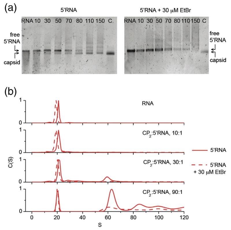
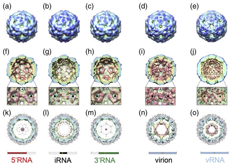
Single-stranded RNA viruses package their genomes into capsids enclosing fixed volumes. We assayed the ability of bacteriophage MS2 coat protein to package large, defined fragments of its genomic, single-stranded RNA. We show that the efficiency of packaging into a T=3 capsid in vitro is inversely proportional to RNA length, implying that there is a free-energy barrier to be overcome during assembly. All the RNAs examined have greater solution persistence lengths than the internal diameter of the capsid into which they become packaged, suggesting that protein-mediated RNA compaction must occur during assembly. Binding ethidium bromide to one of these RNA fragments, which would be expected to reduce its flexibility, severely inhibited packaging, consistent with this idea. Cryo-EM structures of the capsids assembled in these experiments with the sub-genomic RNAs show a layer of RNA density beneath the coat protein shell but lack density for the inner RNA shell seen in the wild-type virion. The inner layer is restored when full-length virion RNA is used in the assembly reaction, implying that it becomes ordered only when the capsid is filled, presumably because of the effects of steric and/or electrostatic repulsions. The cryo-EM results explain the length dependence of packaging. In addition, they show that for the sub-genomic fragments the strongest ordered RNA density occurs below the coat protein dimers forming the icosahedral 5-fold axes of the capsid. There is little such density beneath the proteins at the 2-fold axes, consistent with our model in which coat protein dimers binding to RNA stem-loops located at sites throughout the genome leads to switching of their preferred conformations, thus regulating the placement of the quasi-conformers needed to build the T=3 capsid. The data are consistent with mutual chaperoning of both RNA and coat protein conformations, partially explaining the ability of such viruses to assemble so rapidly and accurately.


Figures

Fig. 1
Fig. 1
The architecture of the T=3 MS2 capsid and sub-genomic RNA fragments. (a) In the T=3 capsid, the MS2 coat protein subunit (CP) is found in three quasi-equivalent conformers: A (blue), B (green) and C (deep red). These conformers form two types of dimer (CP2), an asymmetric A/B dimer, and a symmetric C/C dimer, which differ primarily in the orientation of the loops between the F and G β-strands (FG loop). Conformational switching between symmetric and asymmetric conformations is promoted by the binding of a 19 nt RNA stem–loop (TR). (b) The A/B and C/C dimers are the basic building block of the capsid. The extended FG loops of the A and C conformers pack around the icosahedral 3-fold axis and the compact loops of the B conformer pack around the icosahedral 5-fold axis. (c) Such packing leads to the T=3 caspid, containing 60 A/B and 30 C/C dimers. (d) The RNAs used in this study. The position of the TR assembly-initiation sequence is in gold and boxed, and its sequence is described in (a). The RNAs used for assembly reactions are shown in red (5′RNA), black (iRNA) and green (3′RNA). The full-length genomic RNA extracted from virions is shown in blue. The numbering corresponds to the nucleotide sequence of the genomic RNA (GenBank accession number NC_001417; Table 2). Figures 1 and 5 were produced using UCSF Chimera and PyMOL (www.pymol.org).
Fig. 2
Fig. 2
Gel mobility and electron microscopy assays of MS2 capsid assembly. Assembly reactions with (a) vRNA, (b) 5′RNA, (c) 3′RNA, and (d) iRNA. The top half of (a – d) shows a native agarose gel of capsid assembly reactions induced with the respective RNA. The number above each lane indicates the CP2:RNA stoichiometry; i.e. coat protein dimer:RNA, of the assembly reaction in that lane. The migration positions of the RNA fragment used in each panel, the recombinant MS2 capsid and aggregated material are indicated. Selected reactions were negatively stained with 2% (w/v) uranyl acetate and imaged by electron microscopy. The scale bars represent 200 nm.
Fig. 3
Fig. 3
Assays of packaging efficiency with different lengths of RNA. The figure shows the results of sedimentation velocity assays of the different assembly reactions. The two top-most panels show C(S) versus S plots of capsid assembly reaction components; i.e. coat protein dimer (CP2); T = 3 capsid; vRNA and the unliganded RNA transcripts, all at a concentration of 40 nM. Note, the coat protein dimer sample is at the same concentration as the 90:1 reaction in the lower panel, is in assembly buffer and has been incubated similarly to the RNA–coat protein mixtures; i.e. it is the negative control for the effect of RNA on assembly. Its sedimentation does not differ from coat protein dimer starting material during the course of the experiment. The remaining panels show similar plots for titrations of each RNA with increasing concentrations of CP2 after 4 h at 20 °C in 0.04 M ammonium acetate, 1 mM magnesium acetate, pH 7.2.
Fig. 4
Fig. 4
The effect(s) of ethidium bromide on the efficiency of capsid assembly. Capsid assembly was monitored at increasing stoichiometric ratios of CP2:5′RNA in the presence and in the absence of 30 μM EtBr. (a) Using native agarose gel electrophoresis, no signal corresponding to MS2 capsids was detected in the presence of EtBr after 4 h. (b) Sedimentation velocity analysis confirms that assembly is restricted in the presence of EtBr. 5′ RNA sediments slower in the presence of EtBr, suggesting that EtBr intercalation results in expansion of RNA structure and inhibits folding of the RNA into a structure compatible with the capsid.
Fig. 5
Fig. 5
Cryo-EM structures of MS2 capsids. (a–e) Surface representation of the cryo-EM structures of MS2 capsids reassembled in the presence of 5′RNA (a), iRNA (b), 3′RNA (c) and vRNA (e). For comparison, the cryo-EM structure of the native virion is also shown (d).22 All structures have been Fourier filtered at ~15 Å resolution, and are radially coloured from blue at high radius to red at low radius. (f–j) The rear halves of the 3-D structures of 5′RNA (f), iRNA (g), 3′RNA (h), wt MS2 virion (i) and vRNA (j) with the same radial colouring scheme. The view for each structure is looking down an icosahedral 2-fold axis. The inset is an expanded portion of each structure showing details of the RNA density surrounding the 5-fold axes (indicated by black pentagons). Atomic coordinates (cartoon representation and coloured as in Fig. 1) are fit into a now semi-transparent density. There is little or no density for RNA beneath the C/C dimers (on the 2-fold axes). (k–o) Central, 40 Å thick cross-section views of 5′RNA (k), iRNA (l), 3′RNA (m), the wt MS2 virion (n) and vRNA (o). In each case, the view is along a 3-fold axis, and the density for CP has been masked away. The fitted atomic coordinates are shown as a grey cartoon. The unfiltered density for packaged RNA is coloured radially. The virion (n) shows an outer shell of RNA immediately beneath the protein capsid and a second shell of RNA at lower radii, connected to the outer shell along 5-fold axes. The inner-shell density is not seen in the sub-genomic RNA cryo-EM structures at an equivalent threshold level, and only very weak features are seen in the maps at lower radii (k–m). The inner shell reappears when the vRNA is packaged (o). Schematics for the RNA present in the structures shown in each column are shown for clarity.

Similar articles

Cited by

References

    1. Schneemann A. The structural and functional role of RNA in icosahedral virus assembly. Annu. Rev. Microbiol. 2006;60:51–67. - PubMed
    1. Harrison SC, Olson AJ, Schutt CE, Winkler FK. Tomato bushy stunt virus at 2.9 Å resolution. Nature. 1978;276:368–373. - PubMed
    1. Caspar DL, Klug A. Physical principles in the construction of regular viruses. Cold Spring Harbor Symp. Quant. Biol. 1962;27:1–24. - PubMed
    1. Basnak G, Morton VL, Rolfsson O, Stonehouse NJ, Ashcroft AE, Stockley PG. Viral genomic single-stranded RNA directs the pathway toward a T = 3 capsid. J. Mol. Biol. 2010;395:924–936. - PMC - PubMed
    1. Stockley PG, Rolfsson O, Thompson GS, Basnak G, Francese S, Stonehouse NJ, et al. A simple, RNA-mediated allosteric switch controls the pathway to formation of a T = 3 viral capsid. J. Mol. Biol. 2007;369:541–552. - PMC - PubMed

Publication types

MeSH terms

LinkOut - more resources